3U06 | pdb_00003u06

Crystal structure of the kinesin-14 NcdG347D


Experimental Data Snapshot

  • Method: X-RAY DIFFRACTION
  • Resolution: 2.35 Å
  • R-Value Free: 
    0.250 (Depositor), 0.246 (DCC) 
  • R-Value Work: 
    0.216 (Depositor), 0.217 (DCC) 
  • R-Value Observed: 
    0.217 (Depositor) 

wwPDB Validation 3D Report Full Report

Validation slider image for 3U06

Ligand Structure Quality Assessment 


This is version 1.3 of the entry. See complete history

Literature

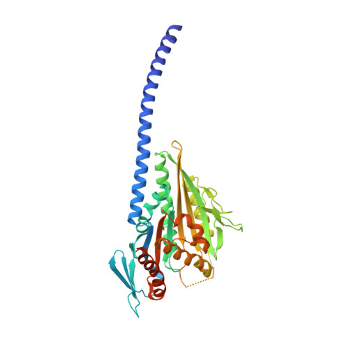
Neck-motor interactions trigger rotation of the kinesin stalk.

Liu, H.L.Pemble Iv, C.W.Endow, S.A.

(2012) Sci Rep 2: 236-236

  • DOI: https://doi.org/10.1038/srep00236
  • Primary Citation Related Structures: 
    3U06

  • PubMed Abstract: 

    Rotation of the coiled-coil stalk of the kinesin-14 motors is thought to drive displacements or steps by the motor along microtubules, but the structural changes that trigger stalk rotation and the nucleotide state in which it occurs are not certain. Here we report a kinesin-14 neck mutant that releases ADP more slowly than wild type and shows weaker microtubule affinity, consistent with defective stalk rotation. Unexpectedly, crystal structures show the stalk fully rotated - neck-motor interactions destabilize the stalk, causing it to rotate and ADP to be released, and alter motor affinity for microtubules. A new structural pathway accounts for the coupling of stalk rotation - the force-producing stroke - to changes in motor affinity for nucleotide and microtubules. Sequential disruption of salt bridges that stabilize the unrotated stalk could cause the stalk to initiate and complete rotation in different nucleotide states.


  • Organizational Affiliation
    • Department of Cell Biology, Duke University Medical Center, Durham, NC 27710 USA.

Macromolecule Content 

  • Total Structure Weight: 94.92 kDa 
  • Atom Count: 5,836 
  • Modeled Residue Count: 703 
  • Deposited Residue Count: 824 
  • Unique protein chains: 1

Macromolecules

Find similar proteins by:|  3D Structure
Entity ID: 1
MoleculeChains  Sequence LengthOrganismDetailsImage
Protein claret segregational
A, B
412Drosophila melanogasterMutation(s): 1 
Gene Names: CA(ND)CG7831ncdnon-claret disjunctional
EC: 3.6.4
UniProt
Find proteins for P20480 (Drosophila melanogaster)
Explore P20480 
Go to UniProtKB:  P20480
Entity Groups
Sequence Clusters30% Identity50% Identity70% Identity90% Identity95% Identity100% Identity
UniProt GroupP20480
Sequence Annotations
Expand
Reference Sequence

Small Molecules

Ligands 3 Unique
IDChains Name / Formula / InChI Key2D Diagram3D Interactions
ADP

Query on ADP


Download:Ideal Coordinates CCD File
C [auth A],
I [auth B]
ADENOSINE-5'-DIPHOSPHATE
C10 H15 N5 O10 P2
XTWYTFMLZFPYCI-KQYNXXCUSA-N
GOL

Query on GOL


Download:Ideal Coordinates CCD File
E [auth A],
F [auth A],
G [auth A],
H [auth A],
J [auth B]
GLYCEROL
C3 H8 O3
PEDCQBHIVMGVHV-UHFFFAOYSA-N
MG

Query on MG


Download:Ideal Coordinates CCD File
D [auth A]MAGNESIUM ION
Mg
JLVVSXFLKOJNIY-UHFFFAOYSA-N

Experimental Data & Validation

Experimental Data

  • Method: X-RAY DIFFRACTION
  • Resolution: 2.35 Å
  • R-Value Free:  0.250 (Depositor), 0.246 (DCC) 
  • R-Value Work:  0.216 (Depositor), 0.217 (DCC) 
  • R-Value Observed: 0.217 (Depositor) 
Space Group: C 1 2 1
Unit Cell:
Length ( Å )Angle ( ˚ )
a = 160.539α = 90
b = 67.094β = 98.91
c = 94.369γ = 90
Software Package:
Software NamePurpose
DENZOdata reduction
SCALEPACKdata scaling
PHASERphasing
PHENIXrefinement
PDB_EXTRACTdata extraction
SERGUIdata collection
HKL-2000data reduction
HKL-2000data scaling

Structure Validation

View Full Validation Report


Ligand Structure Quality Assessment 


Entry History 

Deposition Data

Revision History  (Full details and data files)

  • Version 1.0: 2012-03-07
    Type: Initial release
  • Version 1.1: 2014-11-12
    Changes: Structure summary
  • Version 1.2: 2017-11-08
    Changes: Refinement description
  • Version 1.3: 2024-02-28
    Changes: Data collection, Database references, Derived calculations