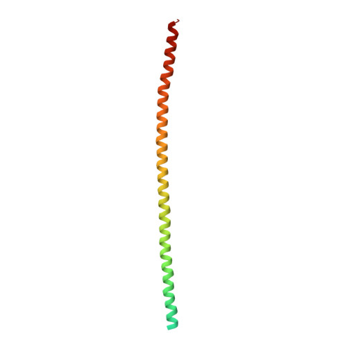
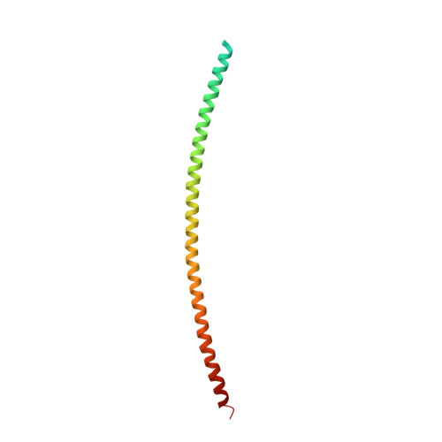
3TNU

Heterocomplex of coil 2B domains of human intermediate filament proteins, keratin 5 (KRT5) and keratin 14 (KRT14)


Experimental Data Snapshot

  • Method: X-RAY DIFFRACTION
  • Resolution: 3.00 Å
  • R-Value Free: 0.234 
  • R-Value Work: 0.209 
  • R-Value Observed: 0.210 

wwPDB Validation   3D Report Full Report


This is version 1.2 of the entry. See complete history


Literature

Structural basis for heteromeric assembly and perinuclear organization of keratin filaments.

Lee, C.H.Kim, M.S.Chung, B.M.Leahy, D.J.Coulombe, P.A.

(2012) Nat Struct Mol Biol 19: 707-715

  • DOI: https://doi.org/10.1038/nsmb.2330
  • Primary Citation of Related Structures:  
    3TNU

  • PubMed Abstract: 

    There is as yet no high-resolution data regarding the structure and organization of keratin intermediate filaments, which are obligate heteropolymers providing vital mechanical support in epithelia. We report the crystal structure of interacting 2B regions from the central coiled-coil domains of keratins 5 and 14 (K5 and K14), expressed in progenitor keratinocytes of epidermis. The interface of the K5-K14 coiled-coil heterodimer has asymmetric salt bridges, hydrogen bonds and hydrophobic contacts, and its surface exhibits a notable charge polarization. A trans-dimer homotypic disulfide bond involving Cys367 in K14's stutter region occurs in the crystal and in skin keratinocytes, where it is concentrated in a keratin filament cage enveloping the nucleus. We show that K14-Cys367 impacts nuclear shape in cultured keratinocytes and that mouse epidermal keratinocytes lacking K14 show aberrations in nuclear structure, highlighting a new function for keratin filaments.


  • Organizational Affiliation

    Department of Biochemistry and Molecular Biology, Bloomberg School of Public Health, Johns Hopkins University, Baltimore, Maryland, USA.


Macromolecules
Find similar proteins by:  (by identity cutoff)  |  3D Structure
Entity ID: 1
MoleculeChains Sequence LengthOrganismDetailsImage
Keratin, type I cytoskeletal 14131Homo sapiensMutation(s): 0 
Gene Names: KRT14
UniProt & NIH Common Fund Data Resources
Find proteins for P02533 (Homo sapiens)
Explore P02533 
Go to UniProtKB:  P02533
PHAROS:  P02533
GTEx:  ENSG00000186847 
Entity Groups  
Sequence Clusters30% Identity50% Identity70% Identity90% Identity95% Identity100% Identity
UniProt GroupP02533
Sequence Annotations
Expand
  • Reference Sequence
Find similar proteins by:  (by identity cutoff)  |  3D Structure
Entity ID: 2
MoleculeChains Sequence LengthOrganismDetailsImage
Keratin, type II cytoskeletal 5129Homo sapiensMutation(s): 0 
Gene Names: KRT5
UniProt & NIH Common Fund Data Resources
Find proteins for P13647 (Homo sapiens)
Explore P13647 
Go to UniProtKB:  P13647
PHAROS:  P13647
GTEx:  ENSG00000186081 
Entity Groups  
Sequence Clusters30% Identity50% Identity70% Identity90% Identity95% Identity100% Identity
UniProt GroupP13647
Sequence Annotations
Expand
  • Reference Sequence
Experimental Data & Validation

Experimental Data

  • Method: X-RAY DIFFRACTION
  • Resolution: 3.00 Å
  • R-Value Free: 0.234 
  • R-Value Work: 0.209 
  • R-Value Observed: 0.210 
  • Space Group: H 3 2
Unit Cell:
Length ( Å )Angle ( ˚ )
a = 150.991α = 90
b = 150.991β = 90
c = 141.623γ = 120
Software Package:
Software NamePurpose
PHENIXrefinement

Structure Validation

View Full Validation Report


Entry History 

Deposition Data

Revision History  (Full details and data files)

  • Version 1.0: 2012-06-20
    Type: Initial release
  • Version 1.1: 2012-08-01
    Changes: Database references
  • Version 1.2: 2024-11-06
    Changes: Data collection, Database references, Structure summary