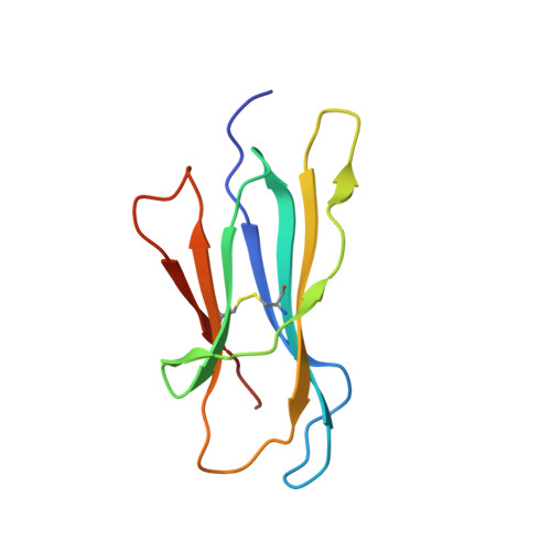
3TM6

Crystal structure of the beta-2 microglobulin DIMC50 disulphide-linked homodimer mutant


Experimental Data Snapshot

  • Method: X-RAY DIFFRACTION
  • Resolution: 2.70 Å
  • R-Value Free: 0.263 
  • R-Value Work: 0.234 
  • R-Value Observed: 0.235 

Starting Model: experimental
View more details

wwPDB Validation   3D Report Full Report


This is version 1.3 of the entry. See complete history


Literature

A recurrent D-strand association interface is observed in beta-2 microglobulin oligomers.

Colombo, M.de Rosa, M.Bellotti, V.Ricagno, S.Bolognesi, M.

(2012) FEBS J 279: 1131-1143

  • DOI: https://doi.org/10.1111/j.1742-4658.2012.08510.x
  • Primary Citation of Related Structures:  
    3TLR, 3TM6

  • PubMed Abstract: 

    β-2 microglobulin (β2m) is an amyloidogenic protein responsible for dialysis-related amyloidosis in man. In the early stages of amyloid fibril formation, β2m associates into dimers and higher oligomers, although the structural details of such aggregates are poorly understood. To characterize the protein-protein interactions supporting the formation of oligomers, three individual β2m cysteine mutants and their disulfide-linked homodimers (DIMC20, DIMC50 and DIMC60) were prepared. Amyloid propensity, oligomerization state in solution and crystallogenesis were tested for each β2m homodimer: DIMC20 and DIMC50 display a mixture of tetrameric and dimeric species in solution and also yield protein crystals and amyloid fibrils, whereas DIMC60 is dimeric in solution but does not form protein crystals nor amyloid fibrils. The X-ray structures of DIMC20 and DIMC50 show that the two engineered dimers form a tetrameric assembly; for both tetrameric species, the noncovalent association interface is based on the interaction of facing β2m D-strands and is conserved. Notably, DIMC20 and DIMC50 trigger amyloid formation in wild-type β2m in unseeded reactions. Thus, when the D-D-strand interface is impaired by an intermolecular disulfide bond (as in DIMC60), the formation of tetramers is hindered, and the protein is not amyloidogenic and does not promote amyloid aggregation of wild-type β2m. Implications for β2m oligomerization are discussed.


  • Organizational Affiliation

    Dipartimento di Scienze Biomolecolari e Biotecnologie and CIMAINA, Università di Milano, Italy.


Macromolecules
Find similar proteins by:  (by identity cutoff)  |  3D Structure
Entity ID: 1
MoleculeChains Sequence LengthOrganismDetailsImage
Beta-2-microglobulin
A, B, C, D, E
A, B, C, D, E, F, G, H
100Homo sapiensMutation(s): 1 
Gene Names: B2MCDABP0092HDCMA22P
UniProt & NIH Common Fund Data Resources
Find proteins for P61769 (Homo sapiens)
Explore P61769 
Go to UniProtKB:  P61769
PHAROS:  P61769
GTEx:  ENSG00000166710 
Entity Groups  
Sequence Clusters30% Identity50% Identity70% Identity90% Identity95% Identity100% Identity
UniProt GroupP61769
Sequence Annotations
Expand
  • Reference Sequence
Small Molecules
Ligands 2 Unique
IDChains Name / Formula / InChI Key2D Diagram3D Interactions
PEG
Query on PEG

Download Ideal Coordinates CCD File 
I [auth A],
J [auth B],
N [auth D],
Q [auth G],
R [auth G]
DI(HYDROXYETHYL)ETHER
C4 H10 O3
MTHSVFCYNBDYFN-UHFFFAOYSA-N
PO4
Query on PO4

Download Ideal Coordinates CCD File 
K [auth B]
L [auth C]
M [auth C]
O [auth E]
P [auth F]
K [auth B],
L [auth C],
M [auth C],
O [auth E],
P [auth F],
S [auth H]
PHOSPHATE ION
O4 P
NBIIXXVUZAFLBC-UHFFFAOYSA-K
Experimental Data & Validation

Experimental Data

  • Method: X-RAY DIFFRACTION
  • Resolution: 2.70 Å
  • R-Value Free: 0.263 
  • R-Value Work: 0.234 
  • R-Value Observed: 0.235 
  • Space Group: P 1 21 1
Unit Cell:
Length ( Å )Angle ( ˚ )
a = 90.102α = 90
b = 56.272β = 115.67
c = 96.374γ = 90
Software Package:
Software NamePurpose
MxCuBEdata collection
MOLREPphasing
BUSTERrefinement
MOSFLMdata reduction
SCALAdata scaling

Structure Validation

View Full Validation Report


Entry History 

Deposition Data

Revision History  (Full details and data files)

  • Version 1.0: 2012-09-05
    Type: Initial release
  • Version 1.1: 2013-08-28
    Changes: Database references
  • Version 1.2: 2023-09-13
    Changes: Data collection, Database references, Derived calculations, Refinement description
  • Version 1.3: 2024-11-06
    Changes: Structure summary