3TL6 | pdb_00003tl6

Crystal structure of purine nucleoside phosphorylase from Entamoeba histolytica


Experimental Data Snapshot

  • Method: X-RAY DIFFRACTION
  • Resolution: 2.65 Å
  • R-Value Free: 
    0.257 (Depositor), 0.248 (DCC) 
  • R-Value Work: 
    0.213 (Depositor) 
  • R-Value Observed: 
    0.215 (Depositor) 

Starting Model: experimental
View more details

wwPDB Validation 3D Report Full Report

Validation slider image for 3TL6

This is version 1.2 of the entry. See complete history

Literature

Expression of proteins in Escherichia coli as fusions with maltose-binding protein to rescue non-expressed targets in a high-throughput protein-expression and purification pipeline.

Hewitt, S.N.Choi, R.Kelley, A.Crowther, G.J.Napuli, A.J.Van Voorhis, W.C.

(2011) Acta Crystallogr Sect F Struct Biol Cryst Commun 67: 1006-1009

  • DOI: https://doi.org/10.1107/S1744309111022159
  • Primary Citation Related Structures: 
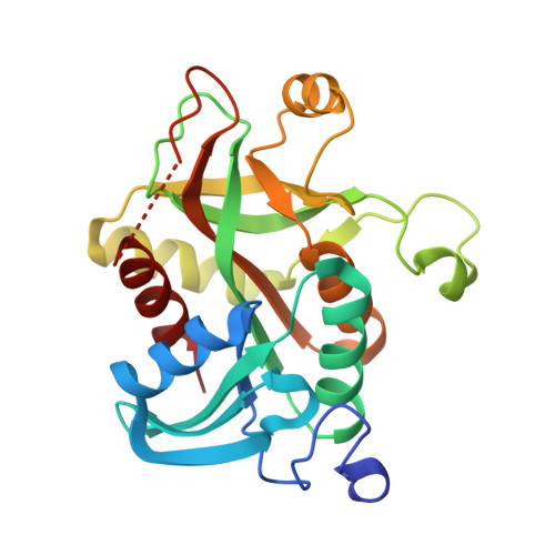
    3SDS, 3TL6, 3U40

  • PubMed Abstract: 

    Despite recent advances, the expression of heterologous proteins in Escherichia coli for crystallization remains a nontrivial challenge. The present study investigates the efficacy of maltose-binding protein (MBP) fusion as a general strategy for rescuing the expression of target proteins. From a group of sequence-verified clones with undetectable levels of protein expression in an E. coli T7 expression system, 95 clones representing 16 phylogenetically diverse organisms were selected for recloning into a chimeric expression vector with an N-terminal histidine-tagged MBP. PCR-amplified inserts were annealed into an identical ligation-independent cloning region in an MBP-fusion vector and were analyzed for expression and solubility by high-throughput nickel-affinity binding. This approach yielded detectable expression of 72% of the clones; soluble expression was visible in 62%. However, the solubility of most proteins was marginal to poor upon cleavage of the MBP tag. This study offers large-scale evidence that MBP can improve the soluble expression of previously non-expressing proteins from a variety of eukaryotic and prokaryotic organisms. While the behavior of the cleaved proteins was disappointing, further refinements in MBP tagging may permit the more widespread use of MBP-fusion proteins in crystallographic studies.


  • Organizational Affiliation
    • Seattle Structural Genomics Center for Infectious Disease (SSGCID), University of Washington, WA 98195, USA.

Macromolecule Content 

  • Total Structure Weight: 160.26 kDa 
  • Atom Count: 10,010 
  • Modeled Residue Count: 1,356 
  • Deposited Residue Count: 1,452 
  • Unique protein chains: 1

Macromolecules

Find similar proteins by:|  3D Structure
Entity ID: 1
MoleculeChains  Sequence LengthOrganismDetailsImage
Purine nucleoside phosphorylase
A, B, C, D, E
A, B, C, D, E, F
242Entamoeba histolyticaMutation(s): 0 
Gene Names: EHI_130960
EC: 2.4.2.1
UniProt
Find proteins for C4LXG4 (Entamoeba histolytica (strain ATCC 30459 / HM-1:IMSS / ABRM))
Explore C4LXG4 
Go to UniProtKB:  C4LXG4
Entity Groups
Sequence Clusters30% Identity50% Identity70% Identity90% Identity95% Identity100% Identity
UniProt GroupC4LXG4
Sequence Annotations
Expand
Reference Sequence

Experimental Data & Validation

Experimental Data

  • Method: X-RAY DIFFRACTION
  • Resolution: 2.65 Å
  • R-Value Free:  0.257 (Depositor), 0.248 (DCC) 
  • R-Value Work:  0.213 (Depositor) 
  • R-Value Observed: 0.215 (Depositor) 
Space Group: P 1 21 1
Unit Cell:
Length ( Å )Angle ( ˚ )
a = 97.97α = 90
b = 86.56β = 117.88
c = 102.56γ = 90
Software Package:
Software NamePurpose
XSCALEdata scaling
PHASERphasing
REFMACrefinement
PDB_EXTRACTdata extraction
ADSCdata collection
XDSdata reduction

Structure Validation

View Full Validation Report


Entry History 

Deposition Data

Revision History  (Full details and data files)

  • Version 1.0: 2011-09-07
    Type: Initial release
  • Version 1.1: 2011-09-21
    Changes: Database references
  • Version 1.2: 2023-09-13
    Changes: Data collection, Database references, Derived calculations, Refinement description