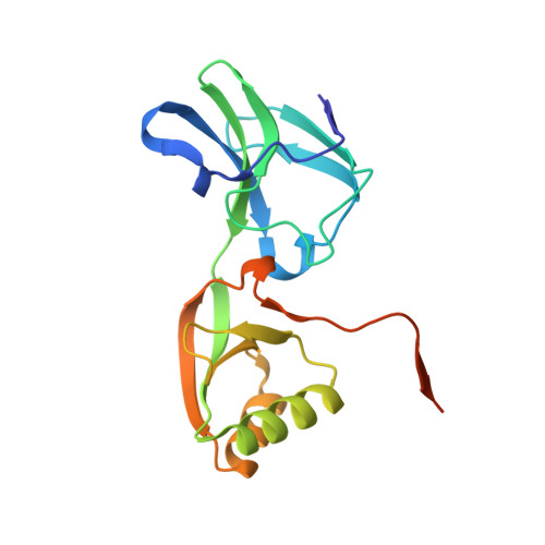
Crystallographic and X-ray absorption spectroscopic characterization of Helicobacter pylori UreE bound to Ni2+ and Zn2+ reveals a role for the disordered C-terminal arm in metal trafficking.
Banaszak, K., Martin-Diaconescu, V., Bellucci, M., Zambelli, B., Rypniewski, W., Maroney, M.J., Ciurli, S.(2012) Biochem J 441: 1017-1026
- PubMed: 22010876
- DOI: https://doi.org/10.1042/BJ20111659
- Primary Citation of Related Structures:
3TJ8, 3TJ9, 3TJA - PubMed Abstract:
The survival and growth of the pathogen Helicobacter pylori in the gastric acidic environment is ensured by the activity of urease, an enzyme containing two essential Ni²⁺ ions in the active site. The metallo-chaperone UreE facilitates in vivo Ni²⁺ insertion into the apoenzyme. Crystals of apo-HpUreE (H. pylori UreE) and its Ni⁺- and Zn⁺-bound forms were obtained from protein solutions in the absence and presence of the metal ions. The crystal structures of the homodimeric protein, determined at 2.00 Å (apo), 1.59 Å (Ni²⁺) and 2.52 Å (Zn²⁺) resolution, show the conserved proximal and solvent-exposed His¹⁰² residues from two adjacent monomers invariably involved in metal binding. The C-terminal regions of the apoprotein are disordered in the crystal, but acquire significant ordering in the presence of the metal ions due to the binding of His¹⁵². The analysis of X-ray absorption spectral data obtained using solutions of Ni²⁺- and Zn²⁺-bound HpUreE provided accurate information of the metal-ion environment in the absence of solid-state effects. These results reveal the role of the histidine residues at the protein C-terminus in metal-ion binding, and the mutual influence of protein framework and metal-ion stereo-electronic properties in establishing co-ordination number and geometry leading to metal selectivity.
- Institute of Bioorganic Chemistry, Polish Academy of Sciences, Noskowskiego 12/14, 61-704 Poznan, Poland.
Organizational Affiliation:


