3TIF | pdb_00003tif

Dimeric structure of a post-hydrolysis state of the ATP-binding cassette MJ0796 bound to ADP and Pi


Experimental Data Snapshot

  • Method: X-RAY DIFFRACTION
  • Resolution: 1.80 Å
  • R-Value Free: 
    0.236 (Depositor), 0.229 (DCC) 
  • R-Value Work: 
    0.203 (Depositor), 0.198 (DCC) 
  • R-Value Observed: 
    0.205 (Depositor) 

Starting Model: experimental
View more details

wwPDB Validation 3D Report Full Report

Validation slider image for 3TIF

Ligand Structure Quality Assessment 


This is version 1.2 of the entry. See complete history

Literature

Kinetics of the association/dissociation cycle of an ATP-binding cassette nucleotide-binding domain.

Zoghbi, M.E.Fuson, K.L.Sutton, R.B.Altenberg, G.A.

(2012) J Biological Chem 287: 4157-4164

  • DOI: https://doi.org/10.1074/jbc.M111.318378
  • Primary Citation Related Structures: 
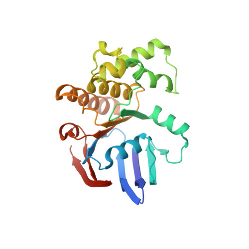
    3TIF

  • PubMed Abstract: 

    Most ATP binding cassette (ABC) proteins are pumps that transport substrates across biological membranes using the energy of ATP hydrolysis. Functional ABC proteins have two nucleotide-binding domains (NBDs) that bind and hydrolyze ATP, but the molecular mechanism of nucleotide hydrolysis is unresolved. This is due in part to the limited kinetic information on NBD association and dissociation. Here, we show dimerization of a catalytically active NBD and follow in real time the association and dissociation of NBDs from the changes in fluorescence emission of a tryptophan strategically located at the center of the dimer interface. Spectroscopic and structural studies demonstrated that the tryptophan can be used as dimerization probe, and we showed that under hydrolysis conditions (millimolar MgATP), not only the dimer dissociation rate increases, but also the dimerization rate. Neither dimer formation or dissociation are clearly favored, and the end result is a dynamic equilibrium where the concentrations of monomer and dimer are very similar. We proposed that based on their variable rates of hydrolysis, the rate-limiting step of the hydrolysis cycle may differ among full-length ABC proteins.


  • Organizational Affiliation
    • Department of Cell Physiology and Molecular Biophysics, Texas Tech Health Sciences Center, Lubbock, Texas 79430-6551, USA.

Macromolecule Content 

  • Total Structure Weight: 54.71 kDa 
  • Atom Count: 3,979 
  • Modeled Residue Count: 462 
  • Deposited Residue Count: 470 
  • Unique protein chains: 1

Macromolecules

Find similar proteins by:|  3D Structure
Entity ID: 1
MoleculeChains  Sequence LengthOrganismDetailsImage
Uncharacterized ABC transporter ATP-binding protein MJ0796
A, B
235Methanocaldococcus jannaschii DSM 2661Mutation(s): 3 
Gene Names: MJ0796
UniProt
Find proteins for Q58206 (Methanocaldococcus jannaschii (strain ATCC 43067 / DSM 2661 / JAL-1 / JCM 10045 / NBRC 100440))
Explore Q58206 
Go to UniProtKB:  Q58206
Entity Groups
Sequence Clusters30% Identity50% Identity70% Identity90% Identity95% Identity100% Identity
UniProt GroupQ58206
Sequence Annotations
Expand
Reference Sequence

Small Molecules

Ligands 4 Unique
IDChains Name / Formula / InChI Key2D Diagram3D Interactions
ADP

Query on ADP


Download:Ideal Coordinates CCD File
C [auth A],
H [auth B]
ADENOSINE-5'-DIPHOSPHATE
C10 H15 N5 O10 P2
XTWYTFMLZFPYCI-KQYNXXCUSA-N
PI

Query on PI


Download:Ideal Coordinates CCD File
D [auth A],
I [auth B]
HYDROGENPHOSPHATE ION
H O4 P
NBIIXXVUZAFLBC-UHFFFAOYSA-L
IPA

Query on IPA


Download:Ideal Coordinates CCD File
E [auth A],
F [auth A],
J [auth B]
ISOPROPYL ALCOHOL
C3 H8 O
KFZMGEQAYNKOFK-UHFFFAOYSA-N
NA

Query on NA


Download:Ideal Coordinates CCD File
G [auth A],
K [auth B]
SODIUM ION
Na
FKNQFGJONOIPTF-UHFFFAOYSA-N

Experimental Data & Validation

Experimental Data

  • Method: X-RAY DIFFRACTION
  • Resolution: 1.80 Å
  • R-Value Free:  0.236 (Depositor), 0.229 (DCC) 
  • R-Value Work:  0.203 (Depositor), 0.198 (DCC) 
  • R-Value Observed: 0.205 (Depositor) 
Space Group: C 2 2 21
Unit Cell:
Length ( Å )Angle ( ˚ )
a = 80.649α = 90
b = 106.347β = 90
c = 117.232γ = 90
Software Package:
Software NamePurpose
ADSCdata collection
PHASERphasing
PHENIXrefinement
HKL-2000data reduction
SCALEPACKdata scaling

Structure Validation

View Full Validation Report


Ligand Structure Quality Assessment 


Entry History 

Deposition Data

  • Released Date: 2012-03-21 
  • Deposition Author(s): Sutton, R.B.

Revision History  (Full details and data files)

  • Version 1.0: 2012-03-21
    Type: Initial release
  • Version 1.1: 2018-03-07
    Changes: Data collection
  • Version 1.2: 2023-09-13
    Changes: Data collection, Database references, Derived calculations, Refinement description