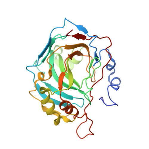
Promiscuity of carbonic anhydrase II. Unexpected ester hydrolysis of carbohydrate-based sulfamate inhibitors.
Lopez, M., Vu, H., Wang, C.K., Wolf, M.G., Groenhof, G., Innocenti, A., Supuran, C.T., Poulsen, S.A.(2011) J Am Chem Soc 133: 18452-18462
- PubMed: 21958118
- DOI: https://doi.org/10.1021/ja207855c
- Primary Citation of Related Structures:
3T82, 3T83, 3T84, 3T85 - PubMed Abstract:
Carbonic anhydrases (CAs) are enzymes whose endogenous reaction is the reversible hydration of CO(2) to give HCO(3)(-) and a proton. CA are also known to exhibit weak and promiscuous esterase activity toward activated esters. Here, we report a series of findings obtained with a set of CA inhibitors that showed quite unexpectedly that the compounds were both inhibitors of CO(2) hydration and substrates for the esterase activity of CA. The compounds comprised a monosaccharide core with the C-6 primary hydroxyl group derivatized as a sulfamate (for CA recognition). The remaining four sugar hydroxyl groups were acylated. Using protein X-ray crystallography, the crystal structures of human CA II in complex with four of the sulfamate inhibitors were obtained. As expected, the four structures displayed the canonical CA protein-sulfamate interactions. Unexpectedly, a free hydroxyl group was observed at the anomeric center (C-1) rather than the parent C-1 acyl group. In addition, this hydroxyl group is observed axial to the carbohydrate ring while in the parent structure it is equatorial. A mechanism is proposed that accounts for this inversion of stereochemistry. For three of the inhibitors, the acyl groups at C-2 or at C-2 and C-3 were also absent with hydroxyl groups observed in their place and retention of stereochemistry. With the use of electrospray ionization-Fourier transform ion cyclotron resonance-mass spectrometry (ESI-FTICR-MS), we observed directly the sequential loss of all four acyl groups from one of the carbohydrate-based sulfamates. For this compound, the inhibitor and substrate binding mode were further analyzed using free energy calculations. These calculations suggested that the parent compound binds almost exclusively as a substrate. To conclude, we have demonstrated that acylated carbohydrate-based sulfamates are simultaneously inhibitor and substrate of human CA II. Our results suggest that, initially, the substrate binding mode dominates, but following hydrolysis, the ligand can also bind as a pure inhibitor thereby competing with the substrate binding mode.
- Eskitis Institute for Cell and Molecular Therapies, Griffith University, Nathan, Queensland 4111, Australia.
Organizational Affiliation:


