3T7A | pdb_00003t7a

Crystal structure of the catalytic domain of human diphosphoinositol pentakisphosphate kinase 2 (PPIP5K2) in complex with ADP at pH 5.2


Experimental Data Snapshot

  • Method: X-RAY DIFFRACTION
  • Resolution: 1.70 Å
  • R-Value Free: 
    0.206 (Depositor), 0.203 (DCC) 
  • R-Value Work: 
    0.179 (Depositor), 0.178 (DCC) 
  • R-Value Observed: 
    0.181 (Depositor) 

Starting Model: experimental
View more details

wwPDB Validation 3D Report Full Report

Validation slider image for 3T7A

Ligand Structure Quality Assessment 


This is version 1.3 of the entry. See complete history

Literature

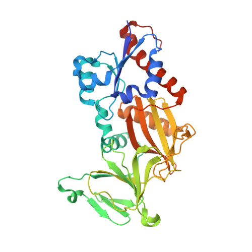
Structural basis for an inositol pyrophosphate kinase surmounting phosphate crowding.

Wang, H.Falck, J.R.Hall, T.M.Shears, S.B.

(2011) Nat Chem Biol 8: 111-116

  • DOI: https://doi.org/10.1038/nchembio.733
  • Primary Citation Related Structures: 
    3T54, 3T7A, 3T99, 3T9A, 3T9B, 3T9C, 3T9D, 3T9E, 3T9F

  • PubMed Abstract: 

    Inositol pyrophosphates (such as IP7 and IP8) are multifunctional signaling molecules that regulate diverse cellular activities. Inositol pyrophosphates have 'high-energy' phosphoanhydride bonds, so their enzymatic synthesis requires that a substantial energy barrier to the transition state be overcome. Additionally, inositol pyrophosphate kinases can show stringent ligand specificity, despite the need to accommodate the steric bulk and intense electronegativity of nature's most concentrated three-dimensional array of phosphate groups. Here we examine how these catalytic challenges are met by describing the structure and reaction cycle of an inositol pyrophosphate kinase at the atomic level. We obtained crystal structures of the kinase domain of human PPIP5K2 complexed with nucleotide cofactors and either substrates, product or a MgF(3)(-) transition-state mimic. We describe the enzyme's conformational dynamics, its unprecedented topological presentation of nucleotide and inositol phosphate, and the charge balance that facilitates partly associative in-line phosphoryl transfer.


  • Organizational Affiliation
    • Inositol Signaling Group, Laboratory of Signal Transduction, National Institute of Environmental Health Sciences, National Institutes of Health, Research Triangle Park, North Carolina, USA. wangh7@niehs.nih.gov

Macromolecule Content 

  • Total Structure Weight: 38.56 kDa 
  • Atom Count: 3,131 
  • Modeled Residue Count: 319 
  • Deposited Residue Count: 330 
  • Unique protein chains: 1

Macromolecules

Find similar proteins by:|  3D Structure
Entity ID: 1
MoleculeChains  Sequence LengthOrganismDetailsImage
Inositol Pyrophosphate Kinase330Homo sapiensMutation(s): 0 
Gene Names: PPIP5K2
EC: 2.7.4.24
UniProt & NIH Common Fund Data Resources
Find proteins for O43314 (Homo sapiens)
Explore O43314 
Go to UniProtKB:  O43314
PHAROS:  O43314
GTEx:  ENSG00000145725 
Entity Groups
Sequence Clusters30% Identity50% Identity70% Identity90% Identity95% Identity100% Identity
UniProt GroupO43314
Sequence Annotations
Expand
Reference Sequence

Small Molecules

Ligands 4 Unique
IDChains Name / Formula / InChI Key2D Diagram3D Interactions
ADP

Query on ADP


Download:Ideal Coordinates CCD File
B [auth A]ADENOSINE-5'-DIPHOSPHATE
C10 H15 N5 O10 P2
XTWYTFMLZFPYCI-KQYNXXCUSA-N
EDO

Query on EDO


Download:Ideal Coordinates CCD File
G [auth A],
I [auth A],
J [auth A],
K [auth A],
M [auth A]
1,2-ETHANEDIOL
C2 H6 O2
LYCAIKOWRPUZTN-UHFFFAOYSA-N
ACT

Query on ACT


Download:Ideal Coordinates CCD File
F [auth A],
H [auth A],
L [auth A]
ACETATE ION
C2 H3 O2
QTBSBXVTEAMEQO-UHFFFAOYSA-M
MG

Query on MG


Download:Ideal Coordinates CCD File
C [auth A],
D [auth A],
E [auth A]
MAGNESIUM ION
Mg
JLVVSXFLKOJNIY-UHFFFAOYSA-N

Experimental Data & Validation

Experimental Data

  • Method: X-RAY DIFFRACTION
  • Resolution: 1.70 Å
  • R-Value Free:  0.206 (Depositor), 0.203 (DCC) 
  • R-Value Work:  0.179 (Depositor), 0.178 (DCC) 
  • R-Value Observed: 0.181 (Depositor) 
Space Group: P 21 21 21
Unit Cell:
Length ( Å )Angle ( ˚ )
a = 88.973α = 90
b = 110.807β = 90
c = 41.381γ = 90
Software Package:
Software NamePurpose
HKL-2000data collection
PHENIXmodel building
REFMACrefinement
HKL-2000data reduction
HKL-2000data scaling
PHENIXphasing

Structure Validation

View Full Validation Report


Ligand Structure Quality Assessment 


Entry History 

Deposition Data

Revision History  (Full details and data files)

  • Version 1.0: 2011-12-07
    Type: Initial release
  • Version 1.1: 2011-12-14
    Changes: Database references
  • Version 1.2: 2012-01-11
    Changes: Database references
  • Version 1.3: 2023-09-13
    Changes: Data collection, Database references, Derived calculations, Refinement description