3T5U | pdb_00003t5u

Crystal structure of the human carbonic anhydrase II in complex with N-hydroxy benzenesulfonamide


Experimental Data Snapshot

  • Method: X-RAY DIFFRACTION
  • Resolution: 1.75 Å
  • R-Value Free: 
    0.183 (Depositor), 0.180 (DCC) 
  • R-Value Work: 
    0.171 (Depositor), 0.170 (DCC) 

Starting Model: experimental
View more details

wwPDB Validation   3D Report Full Report


Ligand Structure Quality Assessment 

Created with Raphaël 2.3.0Worse 01 BetterLigand structure goodness of fit to experimental dataBest fitted MBOClick on this verticalbar to view detailsBest fitted A09Click on this verticalbar to view details

This is version 1.1 of the entry. See complete history


Literature

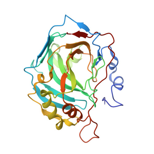
Carbonic anhydrase inhibitors: X-ray crystallographic studies for the binding of N-substituted benzenesulfonamides to human isoform II.

Di Fiore, A.Maresca, A.Alterio, V.Supuran, C.T.De Simone, G.

(2011) Chem Commun (Camb) 47: 11636-11638

  • DOI: https://doi.org/10.1039/c1cc14575d
  • Primary Citation of Related Structures:  
    3T5U, 3T5Z

  • PubMed Abstract: 

    N-substituted benzenesulfonamides, incorporating the N-amino-, N-hydroxy- and N-methoxy-moieties at the sulfonamide zinc binding group, have been investigated as CAIs by means of inhibition and structural studies, unraveling interesting aspects related to their inhibition mechanism.


  • Organizational Affiliation

    Istituto di Biostrutture e Bioimmagini-CNR, via Mezzocannone 16, 80134 Napoli, Italy.


Macromolecules
Find similar proteins by:  (by identity cutoff)  |  3D Structure
Entity ID: 1
MoleculeChains Sequence LengthOrganismDetailsImage
Carbonic anhydrase 2259Homo sapiensMutation(s): 0 
EC: 4.2.1.1 (PDB Primary Data), 4.2.1.69 (UniProt)
UniProt & NIH Common Fund Data Resources
Find proteins for P00918 (Homo sapiens)
Explore P00918 
Go to UniProtKB:  P00918
PHAROS:  P00918
GTEx:  ENSG00000104267 
Entity Groups  
Sequence Clusters30% Identity50% Identity70% Identity90% Identity95% Identity100% Identity
UniProt GroupP00918
Sequence Annotations
Expand
  • Reference Sequence
Small Molecules
Ligands 4 Unique
IDChains Name / Formula / InChI Key2D Diagram3D Interactions
MBO
Query on MBO

Download Ideal Coordinates CCD File 
H [auth A]MERCURIBENZOIC ACID
C7 H5 Hg O2
FVFZSVRSDNUCGG-UHFFFAOYSA-N
A09
Query on A09

Download Ideal Coordinates CCD File 
C [auth A],
D [auth A],
E [auth A],
F [auth A]
N-hydroxybenzenesulfonamide
C6 H7 N O3 S
BRMDATNYMUMZLN-UHFFFAOYSA-N
GOL
Query on GOL

Download Ideal Coordinates CCD File 
G [auth A]GLYCEROL
C3 H8 O3
PEDCQBHIVMGVHV-UHFFFAOYSA-N
ZN
Query on ZN

Download Ideal Coordinates CCD File 
B [auth A]ZINC ION
Zn
PTFCDOFLOPIGGS-UHFFFAOYSA-N
Experimental Data & Validation

Experimental Data

  • Method: X-RAY DIFFRACTION
  • Resolution: 1.75 Å
  • R-Value Free:  0.183 (Depositor), 0.180 (DCC) 
  • R-Value Work:  0.171 (Depositor), 0.170 (DCC) 
Space Group: P 1 21 1
Unit Cell:
Length ( Å )Angle ( ˚ )
a = 42.26α = 90
b = 41.54β = 104.43
c = 72.09γ = 90
Software Package:
Software NamePurpose
CrystalCleardata collection
CNSrefinement
HKL-2000data reduction
HKL-2000data scaling
CNSphasing

Structure Validation

View Full Validation Report


Ligand Structure Quality Assessment 

Created with Raphaël 2.3.0Worse 01 BetterLigand structure goodness of fit to experimental dataBest fitted MBOClick on this verticalbar to view detailsBest fitted A09Click on this verticalbar to view details

Entry History 

Deposition Data

Revision History  (Full details and data files)

  • Version 1.0: 2012-06-20
    Type: Initial release
  • Version 1.1: 2023-09-13
    Changes: Data collection, Database references, Derived calculations, Refinement description