3T4R | pdb_00003t4r

Lettuce Necrotic Yellow Virus Phosphoprotein C-Terminal Domain


Experimental Data Snapshot

  • Method: X-RAY DIFFRACTION
  • Resolution: 2.00 Å
  • R-Value Free: 
    0.294 (Depositor), 0.294 (DCC) 
  • R-Value Work: 
    0.226 (Depositor), 0.226 (DCC) 
  • R-Value Observed: 
    0.229 (Depositor) 

wwPDB Validation 3D Report Full Report

Validation slider image for 3T4R

This is version 1.2 of the entry. See complete history

Literature

Structure of the C-terminal domain of lettuce necrotic yellows virus phosphoprotein.

Martinez, N.Ribeiro, E.A.Leyrat, C.Tarbouriech, N.Ruigrok, R.W.Jamin, M.

(2013) J Virol 87: 9569-9578

  • DOI: https://doi.org/10.1128/JVI.00999-13
  • Primary Citation Related Structures: 
    3T4R

  • PubMed Abstract: 

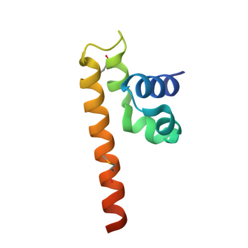
    Lettuce necrotic yellows virus (LNYV) is a prototype of the plant-adapted cytorhabdoviruses. Through a meta-prediction of disorder, we localized a folded C-terminal domain in the amino acid sequence of its phosphoprotein. This domain consists of an autonomous folding unit that is monomeric in solution. Its structure, solved by X-ray crystallography, reveals a lollipop-shaped structure comprising five helices. The structure is different from that of the corresponding domains of other Rhabdoviridae, Filoviridae, and Paramyxovirinae; only the overall topology of the polypeptide chain seems to be conserved, suggesting that this domain evolved under weak selective pressure and varied in size by the acquisition or loss of functional modules.


  • Organizational Affiliation
    • Université Grenoble Alpes, UVHCI, Grenoble, France.

Macromolecule Content 

  • Total Structure Weight: 9.58 kDa 
  • Atom Count: 610 
  • Modeled Residue Count: 72 
  • Deposited Residue Count: 81 
  • Unique protein chains: 1

Macromolecules

Find similar proteins by:|  3D Structure
Entity ID: 1
MoleculeChains  Sequence LengthOrganismDetailsImage
Phosphoprotein81Lettuce necrotic yellows virus isolate 318Mutation(s): 0 
Gene Names: P
UniProt
Find proteins for Q9E7N7 (Lettuce necrotic yellows virus (isolate 318))
Explore Q9E7N7 
Go to UniProtKB:  Q9E7N7
Entity Groups
Sequence Clusters30% Identity50% Identity70% Identity90% Identity95% Identity100% Identity
UniProt GroupQ9E7N7
Sequence Annotations
Expand
Reference Sequence

Small Molecules

Ligands 1 Unique
IDChains Name / Formula / InChI Key2D Diagram3D Interactions
MG

Query on MG


Download:Ideal Coordinates CCD File
B [auth A]MAGNESIUM ION
Mg
JLVVSXFLKOJNIY-UHFFFAOYSA-N
Modified Residues  1 Unique
IDChains TypeFormula2D DiagramParent
MSE
Query on MSE
A
L-PEPTIDE LINKINGC5 H11 N O2 SeMET

Experimental Data & Validation

Experimental Data

  • Method: X-RAY DIFFRACTION
  • Resolution: 2.00 Å
  • R-Value Free:  0.294 (Depositor), 0.294 (DCC) 
  • R-Value Work:  0.226 (Depositor), 0.226 (DCC) 
  • R-Value Observed: 0.229 (Depositor) 
Space Group: P 41 2 2
Unit Cell:
Length ( Å )Angle ( ˚ )
a = 43.28α = 90
b = 43.28β = 90
c = 89.38γ = 90
Software Package:
Software NamePurpose
DNAdata collection
SHELXDphasing
REFMACrefinement
XDSdata reduction
XSCALEdata scaling

Structure Validation

View Full Validation Report


Entry History 

Deposition Data

Revision History  (Full details and data files)

  • Version 1.0: 2013-01-30
    Type: Initial release
  • Version 1.1: 2014-03-26
    Changes: Database references
  • Version 1.2: 2024-10-09
    Changes: Data collection, Database references, Derived calculations, Structure summary