3T30 | pdb_00003t30

Human nucleoplasmin (Npm2): a histone chaperone in oocytes and early embryos


Experimental Data Snapshot

  • Method: X-RAY DIFFRACTION
  • Resolution: 1.90 Å
  • R-Value Free: 
    0.243 (Depositor), 0.247 (DCC) 
  • R-Value Work: 
    0.193 (Depositor), 0.206 (DCC) 
  • R-Value Observed: 
    0.193 (Depositor) 

Starting Model: experimental
View more details

wwPDB Validation 3D Report Full Report

Validation slider image for 3T30

This is version 1.3 of the entry. See complete history

Literature

Crystal structure and function of human nucleoplasmin (npm2): a histone chaperone in oocytes and embryos.

Platonova, O.Akey, I.V.Head, J.F.Akey, C.W.

(2011) Biochemistry 50: 8078-8089

  • DOI: https://doi.org/10.1021/bi2006652
  • Primary Citation Related Structures: 
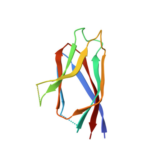
    3T30

  • PubMed Abstract: 

    Human Npm2 is an ortholog of Xenopus nucleoplasmin (Np), a chaperone that binds histones. We have determined the crystal structure of a truncated Npm2-core at 1.9 Å resolution and show that the N-terminal domains of Npm2 and Np form similar pentamers. This allowed us to model an Npm2 decamer which may be formed by hydrogen bonds between quasi-conserved residues in the interface between two pentamers. Interestingly, the Npm2 pentamer lacks a prototypical A1-acidic tract in each of its subunits. This feature may be responsible for the inability of Npm2-core to bind histones. However, Npm2 with a large acidic tract in its C-terminal tail (Npm2-A2) is able to bind histones and form large complexes. Fluorescence resonance energy transfer experiments and biochemical analysis of loop mutations support the premise that nucleoplasmins form decamers when they bind H2A-H2B dimers and H3-H4 tetramers simultaneously. In the absence of histone tetramers, these chaperones bind H2A-H2B dimers with a single pentamer forming the central hub. When taken together, our data provide insights into the mechanism of histone binding by nucleoplasmins.


  • Organizational Affiliation
    • Department of Physiology and Biophysics, Boston University School of Medicine, 700 Albany St., Boston, Massachusetts 02118-2526, USA.

Macromolecule Content 

  • Total Structure Weight: 123.94 kDa 
  • Atom Count: 7,862 
  • Modeled Residue Count: 922 
  • Deposited Residue Count: 1,100 
  • Unique protein chains: 1

Macromolecules

Find similar proteins by:|  3D Structure
Entity ID: 1
MoleculeChains  Sequence LengthOrganismDetailsImage
Nucleoplasmin-2110Homo sapiensMutation(s): 0 
Gene Names: NPM2
UniProt & NIH Common Fund Data Resources
Find proteins for Q86SE8 (Homo sapiens)
Explore Q86SE8 
Go to UniProtKB:  Q86SE8
PHAROS:  Q86SE8
GTEx:  ENSG00000158806 
Entity Groups
Sequence Clusters30% Identity50% Identity70% Identity90% Identity95% Identity100% Identity
UniProt GroupQ86SE8
Sequence Annotations
Expand
Reference Sequence

Experimental Data & Validation

Experimental Data

  • Method: X-RAY DIFFRACTION
  • Resolution: 1.90 Å
  • R-Value Free:  0.243 (Depositor), 0.247 (DCC) 
  • R-Value Work:  0.193 (Depositor), 0.206 (DCC) 
  • R-Value Observed: 0.193 (Depositor) 
Space Group: P 1 21 1
Unit Cell:
Length ( Å )Angle ( ˚ )
a = 49.03α = 90
b = 107.24β = 102.54
c = 108.7γ = 90
Software Package:
Software NamePurpose
PHENIXrefinement
CNSrefinement
ADSCdata collection
DENZOdata reduction
SCALEPACKdata scaling
CNSphasing

Structure Validation

View Full Validation Report


Entry History 

Deposition Data

Revision History  (Full details and data files)

  • Version 1.0: 2011-09-21
    Type: Initial release
  • Version 1.1: 2011-11-30
    Changes: Database references
  • Version 1.2: 2019-07-17
    Changes: Data collection, Refinement description
  • Version 1.3: 2023-09-13
    Changes: Data collection, Database references, Refinement description