3T1F | pdb_00003t1f

Crystal structure of the mouse CD1d-Glc-DAG-s2 complex


Experimental Data Snapshot

  • Method: X-RAY DIFFRACTION
  • Resolution: 1.70 Å
  • R-Value Free: 
    0.219 (Depositor), 0.243 (DCC) 
  • R-Value Work: 
    0.187 (Depositor), 0.220 (DCC) 
  • R-Value Observed: 
    0.188 (Depositor) 

wwPDB Validation 3D Report Full Report

Validation slider image for 3T1F

Ligand Structure Quality Assessment 


This is version 2.1 of the entry. See complete history

Literature

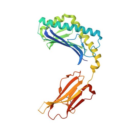
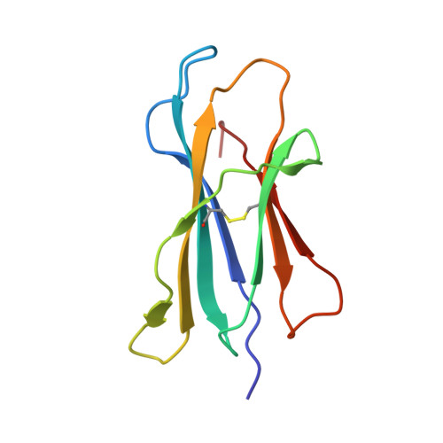
Invariant natural killer T cells recognize glycolipids from pathogenic Gram-positive bacteria.

Kinjo, Y.Illarionov, P.Vela, J.L.Pei, B.Girardi, E.Li, X.Li, Y.Imamura, M.Kaneko, Y.Okawara, A.Miyazaki, Y.Gomez-Velasco, A.Rogers, P.Dahesh, S.Uchiyama, S.Khurana, A.Kawahara, K.Yesilkaya, H.Andrew, P.W.Wong, C.H.Kawakami, K.Nizet, V.Besra, G.S.Tsuji, M.Zajonc, D.M.Kronenberg, M.

(2011) Nat Immunol 12: 966-974

  • DOI: https://doi.org/10.1038/ni.2096
  • Primary Citation Related Structures: 
    3T1F

  • PubMed Abstract: 

    Natural killer T cells (NKT cells) recognize glycolipid antigens presented by CD1d. These cells express an evolutionarily conserved, invariant T cell antigen receptor (TCR), but the forces that drive TCR conservation have remained uncertain. Here we show that NKT cells recognized diacylglycerol-containing glycolipids from Streptococcus pneumoniae, the leading cause of community-acquired pneumonia, and group B Streptococcus, which causes neonatal sepsis and meningitis. Furthermore, CD1d-dependent responses by NKT cells were required for activation and host protection. The glycolipid response was dependent on vaccenic acid, which is present in low concentrations in mammalian cells. Our results show how microbial lipids position the sugar for recognition by the invariant TCR and, most notably, extend the range of microbes recognized by this conserved TCR to several clinically important bacteria.


  • Organizational Affiliation
    • Division of Developmental Immunology, La Jolla Institute for Allergy & Immunology, La Jolla, California, USA.

Macromolecule Content 

  • Total Structure Weight: 46.38 kDa 
  • Atom Count: 3,657 
  • Modeled Residue Count: 362 
  • Deposited Residue Count: 384 
  • Unique protein chains: 2

Macromolecules

Find similar proteins by:|  3D Structure
Entity ID: 1
MoleculeChains  Sequence LengthOrganismDetailsImage
Antigen-presenting glycoprotein CD1d1285Mus musculusMutation(s): 1 
Gene Names: Cd1.1Cd1d1
UniProt & NIH Common Fund Data Resources
Find proteins for P11609 (Mus musculus)
Explore P11609 
Go to UniProtKB:  P11609
IMPC:  MGI:107674
Entity Groups
Sequence Clusters30% Identity50% Identity70% Identity90% Identity95% Identity100% Identity
UniProt GroupP11609
Glycosylation
Glycosylation Sites: 3Go to GlyGen: P11609-1
Sequence Annotations
Expand
Reference Sequence
Find similar proteins by:|  3D Structure
Entity ID: 2
MoleculeChains  Sequence LengthOrganismDetailsImage
Beta-2-microglobulin99Mus musculusMutation(s): 1 
Gene Names: B2mbeta-2-microglobulin
UniProt & NIH Common Fund Data Resources
Find proteins for P01887 (Mus musculus)
Explore P01887 
Go to UniProtKB:  P01887
IMPC:  MGI:88127
Entity Groups
Sequence Clusters30% Identity50% Identity70% Identity90% Identity95% Identity100% Identity
UniProt GroupP01887
Sequence Annotations
Expand
Reference Sequence

Oligosaccharides

Help  
Entity ID: 3
MoleculeChains Length2D Diagram GlycosylationD Interactions
2-acetamido-2-deoxy-beta-D-glucopyranose-(1-4)-2-acetamido-2-deoxy-beta-D-glucopyranose
C
2N-Glycosylation
Glycosylation Resources
GlyTouCan: G42666HT
GlyCosmos: G42666HT
GlyGen: G42666HT

Small Molecules

Ligands 3 Unique
IDChains Name / Formula / InChI Key2D Diagram3D Interactions
3TF

Query on 3TF


Download:Ideal Coordinates CCD File
F [auth A](2S)-1-(alpha-D-glucopyranosyloxy)-3-(hexadecanoyloxy)propan-2-yl (11Z)-octadec-11-enoate
C43 H80 O10
FJAAFGBZVPKOCR-HBHGLPGMSA-N
NAG

Query on NAG


Download:Ideal Coordinates CCD File
D [auth A],
E [auth A]
2-acetamido-2-deoxy-beta-D-glucopyranose
C8 H15 N O6
OVRNDRQMDRJTHS-FMDGEEDCSA-N
GOL

Query on GOL


Download:Ideal Coordinates CCD File
G [auth A],
H [auth A],
I [auth A],
J [auth A],
K [auth A]
GLYCEROL
C3 H8 O3
PEDCQBHIVMGVHV-UHFFFAOYSA-N

Experimental Data & Validation

Experimental Data

  • Method: X-RAY DIFFRACTION
  • Resolution: 1.70 Å
  • R-Value Free:  0.219 (Depositor), 0.243 (DCC) 
  • R-Value Work:  0.187 (Depositor), 0.220 (DCC) 
  • R-Value Observed: 0.188 (Depositor) 
Space Group: P 21 21 21
Unit Cell:
Length ( Å )Angle ( ˚ )
a = 42.191α = 90
b = 107.381β = 90
c = 110.305γ = 90
Software Package:
Software NamePurpose
Blu-Icedata collection
MOLREPphasing
REFMACrefinement
MOSFLMdata reduction
SCALAdata scaling

Structure Validation

View Full Validation Report


Ligand Structure Quality Assessment 


Entry History 

Deposition Data

Revision History  (Full details and data files)

  • Version 1.0: 2011-09-21
    Type: Initial release
  • Version 1.1: 2011-10-05
    Changes: Database references
  • Version 2.0: 2020-07-29
    Type: Remediation
    Reason: Carbohydrate remediation
    Changes: Advisory, Atomic model, Data collection, Database references, Derived calculations, Source and taxonomy, Structure summary
  • Version 2.1: 2024-11-27
    Changes: Advisory, Data collection, Database references, Structure summary