3SXU | pdb_00003sxu

Structure of the E. coli SSB-DNA polymerase III interface


Experimental Data Snapshot

  • Method: X-RAY DIFFRACTION
  • Resolution: 1.85 Å
  • R-Value Free: 
    0.208 (Depositor), 0.208 (DCC) 
  • R-Value Work: 
    0.184 (Depositor), 0.182 (DCC) 
  • R-Value Observed: 
    0.186 (Depositor) 

Starting Model: experimental
View more details

wwPDB Validation 3D Report Full Report

Validation slider image for 3SXU

This is version 1.2 of the entry. See complete history

Literature

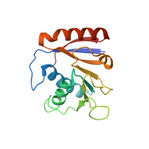
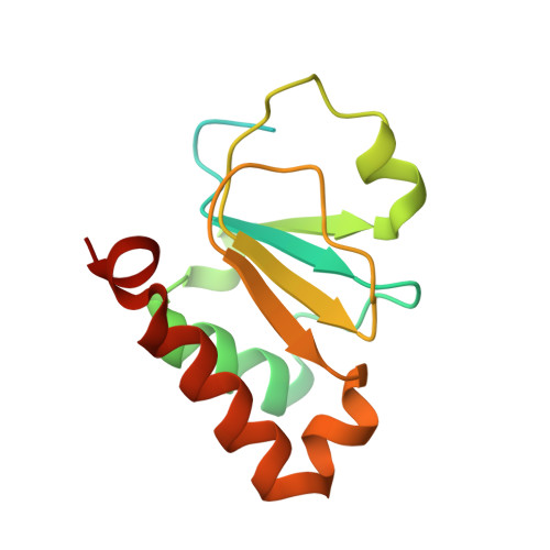
Structure of the SSB-DNA polymerase III interface and its role in DNA replication.

Marceau, A.H.Bahng, S.Massoni, S.C.George, N.P.Sandler, S.J.Marians, K.J.Keck, J.L.

(2011) EMBO J 30: 4236-4247

  • DOI: https://doi.org/10.1038/emboj.2011.305
  • Primary Citation Related Structures: 
    3SXU

  • PubMed Abstract: 

    Interactions between single-stranded DNA-binding proteins (SSBs) and the DNA replication machinery are found in all organisms, but the roles of these contacts remain poorly defined. In Escherichia coli, SSB's association with the χ subunit of the DNA polymerase III holoenzyme has been proposed to confer stability to the replisome and to aid delivery of primers to the lagging-strand DNA polymerase. Here, the SSB-binding site on χ is identified crystallographically and biochemical and cellular studies delineate the consequences of destabilizing the χ/SSB interface. An essential role for the χ/SSB interaction in lagging-strand primer utilization is not supported. However, sequence changes in χ that block complex formation with SSB lead to salt-dependent uncoupling of leading- and lagging-strand DNA synthesis and to a surprising obstruction of the leading-strand DNA polymerase in vitro, pointing to roles for the χ/SSB complex in replisome establishment and maintenance. Destabilization of the χ/SSB complex in vivo produces cells with temperature-dependent cell cycle defects that appear to arise from replisome instability.


  • Organizational Affiliation
    • Department of Biomolecular Chemistry, University of Wisconsin School of Medicine and Public Health, Madison, WI 53706-1532, USA.

Macromolecule Content 

  • Total Structure Weight: 32.86 kDa 
  • Atom Count: 2,349 
  • Modeled Residue Count: 252 
  • Deposited Residue Count: 293 
  • Unique protein chains: 3

Macromolecules

Find similar proteins by:|  3D Structure
Entity ID: 1
MoleculeChains  Sequence LengthOrganismDetailsImage
DNA polymerase III subunit chi150Escherichia coli K-12Mutation(s): 0 
Gene Names: b4259holCJW4216
EC: 2.7.7.7
UniProt
Find proteins for P28905 (Escherichia coli (strain K12))
Explore P28905 
Go to UniProtKB:  P28905
Entity Groups
Sequence Clusters30% Identity50% Identity70% Identity90% Identity95% Identity100% Identity
UniProt GroupP28905
Sequence Annotations
Expand
Reference Sequence
Find similar proteins by:|  3D Structure
Entity ID: 2
MoleculeChains  Sequence LengthOrganismDetailsImage
DNA polymerase III subunit psi138Escherichia coli K-12Mutation(s): 0 
Gene Names: b4372holDJW4334
EC: 2.7.7.7
UniProt
Find proteins for P28632 (Escherichia coli (strain K12))
Explore P28632 
Go to UniProtKB:  P28632
Entity Groups
Sequence Clusters30% Identity50% Identity70% Identity90% Identity95% Identity100% Identity
UniProt GroupP28632
Sequence Annotations
Expand
Reference Sequence
Find similar proteins by:  Sequence   |   3D Structure  
Entity ID: 3
MoleculeChains  Sequence LengthOrganismDetailsImage
SSB peptide5Escherichia coli K-12Mutation(s): 0 
UniProt
Find proteins for P0AGE0 (Escherichia coli (strain K12))
Explore P0AGE0 
Go to UniProtKB:  P0AGE0
Entity Groups
UniProt GroupP0AGE0
Sequence Annotations
Expand
Reference Sequence

Experimental Data & Validation

Experimental Data

  • Method: X-RAY DIFFRACTION
  • Resolution: 1.85 Å
  • R-Value Free:  0.208 (Depositor), 0.208 (DCC) 
  • R-Value Work:  0.184 (Depositor), 0.182 (DCC) 
  • R-Value Observed: 0.186 (Depositor) 
Space Group: P 61 2 2
Unit Cell:
Length ( Å )Angle ( ˚ )
a = 79.927α = 90
b = 79.927β = 90
c = 155.045γ = 120
Software Package:
Software NamePurpose
ADSCdata collection
PHASERphasing
REFMACrefinement
HKL-2000data reduction
HKL-2000data scaling

Structure Validation

View Full Validation Report


Entry History 

Deposition Data

Revision History  (Full details and data files)

  • Version 1.0: 2011-09-21
    Type: Initial release
  • Version 1.1: 2011-11-30
    Changes: Database references
  • Version 1.2: 2023-09-13
    Changes: Data collection, Database references, Refinement description