3SUR | pdb_00003sur

Crystal structure of beta-hexosaminidase from Paenibacillus sp. TS12 in complex with NAG-thiazoline.


Experimental Data Snapshot

  • Method: X-RAY DIFFRACTION
  • Resolution: 1.90 Å
  • R-Value Free: 
    0.181 (Depositor), 0.181 (DCC) 
  • R-Value Work: 
    0.140 (Depositor), 0.141 (DCC) 
  • R-Value Observed: 
    0.142 (Depositor) 

Starting Model: experimental
View more details

wwPDB Validation 3D Report Full Report

Validation slider image for 3SUR

Ligand Structure Quality Assessment 


This is version 1.2 of the entry. See complete history

Literature

Gaining insight into the inhibition of glycoside hydrolase family 20 exo-beta-N-acetylhexosaminidases using a structural approach

Sumida, T.Stubbs, K.A.Ito, M.Yokoyama, S.

(2012) Org Biomol Chem 10: 2607-2612

  • DOI: https://doi.org/10.1039/c2ob06636j
  • Primary Citation Related Structures: 
    3SUR, 3SUS, 3SUT, 3SUU, 3SUV, 3SUW

  • PubMed Abstract: 

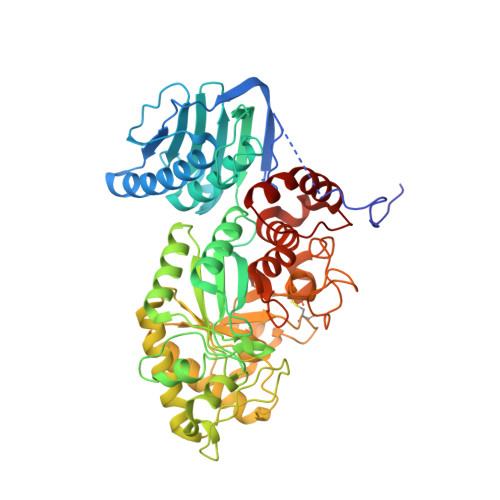
    One useful methodology that has been used to give insight into how chemically synthesized inhibitors bind to enzymes and the reasons underlying their potency is crystallographic studies of inhibitor-enzyme complexes. Presented here is the X-ray structural analysis of a representative family 20 exo-β-N-acetylhexosaminidase in complex with various known classes of inhibitor of these types of enzymes, which highlights how different inhibitor classes can inhibit the same enzyme. This study will aid in the future development of inhibitors of not only exo-β-N-acetylhexosaminidases but also other types of glycoside hydrolases.


  • Organizational Affiliation
    • RIKEN Systems and Structural Biology Center, 1-7-22 Suehiro-cho, Tsurumi, Yokohama 230-0045, Japan.

Macromolecule Content 

  • Total Structure Weight: 57.88 kDa 
  • Atom Count: 4,554 
  • Modeled Residue Count: 504 
  • Deposited Residue Count: 525 
  • Unique protein chains: 1

Macromolecules

Find similar proteins by:|  3D Structure
Entity ID: 1
MoleculeChains  Sequence LengthOrganismDetailsImage
Beta-hexosaminidase525Paenibacillus sp. TS12Mutation(s): 0 
Gene Names: Hex1
EC: 3.2.1.52
UniProt
Find proteins for D2KW09 (Paenibacillus sp. TS12)
Explore D2KW09 
Go to UniProtKB:  D2KW09
Entity Groups
Sequence Clusters30% Identity50% Identity70% Identity90% Identity95% Identity100% Identity
UniProt GroupD2KW09
Sequence Annotations
Expand
Reference Sequence

Small Molecules

Ligands 2 Unique
IDChains Name / Formula / InChI Key2D Diagram3D Interactions
NGT

Query on NGT


Download:Ideal Coordinates CCD File
C [auth A]3AR,5R,6S,7R,7AR-5-HYDROXYMETHYL-2-METHYL-5,6,7,7A-TETRAHYDRO-3AH-PYRANO[3,2-D]THIAZOLE-6,7-DIOL
C8 H13 N O4 S
DRHXTSWSUAJOJZ-FMDGEEDCSA-N
SO4

Query on SO4


Download:Ideal Coordinates CCD File
B [auth A]SULFATE ION
O4 S
QAOWNCQODCNURD-UHFFFAOYSA-L

Experimental Data & Validation

Experimental Data

  • Method: X-RAY DIFFRACTION
  • Resolution: 1.90 Å
  • R-Value Free:  0.181 (Depositor), 0.181 (DCC) 
  • R-Value Work:  0.140 (Depositor), 0.141 (DCC) 
  • R-Value Observed: 0.142 (Depositor) 
Space Group: C 2 2 21
Unit Cell:
Length ( Å )Angle ( ˚ )
a = 95.696α = 90
b = 102.094β = 90
c = 108.06γ = 90
Software Package:
Software NamePurpose
HKL-2000data collection
MOLREPphasing
REFMACrefinement
HKL-2000data reduction
SCALEPACKdata scaling

Structure Validation

View Full Validation Report


Ligand Structure Quality Assessment 


Entry History 

Deposition Data

Revision History  (Full details and data files)

  • Version 1.0: 2012-06-06
    Type: Initial release
  • Version 1.1: 2023-11-01
    Changes: Data collection, Database references, Derived calculations, Refinement description
  • Version 1.2: 2024-10-09
    Changes: Structure summary