3SPF | pdb_00003spf

Crystal Structure of Bcl-xL bound to BM501


Experimental Data Snapshot

  • Method: X-RAY DIFFRACTION
  • Resolution: 1.70 Å
  • R-Value Free: 
    0.204 (Depositor), 0.198 (DCC) 
  • R-Value Work: 
    0.184 (Depositor), 0.182 (DCC) 
  • R-Value Observed: 
    0.185 (Depositor) 

wwPDB Validation 3D Report Full Report

Validation slider image for 3SPF

Ligand Structure Quality Assessment 


This is version 2.2 of the entry. See complete history

Literature

Design of Bcl-2 and Bcl-xL Inhibitors with Subnanomolar Binding Affinities Based upon a New Scaffold.

Zhou, H.Chen, J.Meagher, J.L.Yang, C.Y.Aguilar, A.Liu, L.Bai, L.Cong, X.Cai, Q.Fang, X.Stuckey, J.A.Wang, S.

(2012) J Med Chem 55: 4664-4682

  • DOI: https://doi.org/10.1021/jm300178u
  • Primary Citation Related Structures: 
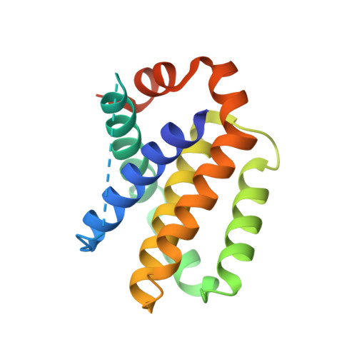
    3SPF

  • PubMed Abstract: 

    Employing a structure-based strategy, we have designed a new class of potent small-molecule inhibitors of the anti-apoptotic proteins Bcl-2 and Bcl-xL. An initial lead compound with a new scaffold was designed based upon the crystal structure of Bcl-xL and U.S. Food and Drug Administration (FDA) approved drugs and was found to have an affinity of 100 μM for both Bcl-2 and Bcl-xL. Linking this weak lead to another weak-affinity fragment derived from Abbott's ABT-737 led to an improvement of the binding affinity by a factor of >10 000. Further optimization ultimately yielded compounds with subnanomolar binding affinities for both Bcl-2 and Bcl-xL and potent cellular activity. The best compound (21) binds to Bcl-xL and Bcl-2 with K(i) < 1 nM, inhibits cell growth in the H146 and H1417 small-cell lung cancer cell lines with IC(50) values of 60-90 nM, and induces robust cell death in the H146 cancer cell line at 30-100 nM.


  • Organizational Affiliation
    • Comprehensive Cancer Center and Department of Internal Medicine, University of Michigan , 1500 East Medical Center Drive, Ann Arbor, Michigan 48109-0934, United States.

Macromolecule Content 

  • Total Structure Weight: 20.57 kDa 
  • Atom Count: 1,378 
  • Modeled Residue Count: 142 
  • Deposited Residue Count: 172 
  • Unique protein chains: 1

Macromolecules

Find similar proteins by:|  3D Structure
Entity ID: 1
MoleculeChains  Sequence LengthOrganismDetailsImage
Bcl-2-like protein 1172Homo sapiensMutation(s): 0 
Gene Names: BCL2L1BCL2LBCLX
UniProt & NIH Common Fund Data Resources
Find proteins for Q07817 (Homo sapiens)
Explore Q07817 
Go to UniProtKB:  Q07817
PHAROS:  Q07817
GTEx:  ENSG00000171552 
Entity Groups
Sequence Clusters30% Identity50% Identity70% Identity90% Identity95% Identity100% Identity
UniProt GroupQ07817
Sequence Annotations
Expand
Reference Sequence

Experimental Data & Validation

Experimental Data

  • Method: X-RAY DIFFRACTION
  • Resolution: 1.70 Å
  • R-Value Free:  0.204 (Depositor), 0.198 (DCC) 
  • R-Value Work:  0.184 (Depositor), 0.182 (DCC) 
  • R-Value Observed: 0.185 (Depositor) 
Space Group: P 42 21 2
Unit Cell:
Length ( Å )Angle ( ˚ )
a = 103.748α = 90
b = 103.748β = 90
c = 35.32γ = 90
Software Package:
Software NamePurpose
DENZOdata reduction
SCALEPACKdata scaling
PHASERphasing
BUSTER-TNTrefinement
PDB_EXTRACTdata extraction
MD2data collection
HKL-2000data reduction
BUSTERrefinement

Structure Validation

View Full Validation Report


Ligand Structure Quality Assessment 


Entry History 

Deposition Data

Revision History  (Full details and data files)

  • Version 1.0: 2012-06-27
    Type: Initial release
  • Version 2.0: 2017-06-28
    Changes: Advisory, Database references, Polymer sequence, Source and taxonomy, Structure summary
  • Version 2.1: 2017-11-08
    Changes: Refinement description
  • Version 2.2: 2024-02-28
    Changes: Advisory, Data collection, Database references, Derived calculations