


Crystal structure of BA2930 in complex with AcCoA and uracil
Klimecka, M.M., Chruszcz, M., Porebski, P.J., Cymborowski, M., Anderson, W., Minor, W., Center for Structural Genomics of Infectious Diseases (CSGID)To be published.
Experimental Data Snapshot
Starting Model: experimental
View more details
Entity ID: 1 | |||||
|---|---|---|---|---|---|
| Molecule | Chains | Sequence Length | Organism | Details | Image |
| Aminoglycoside N3-acetyltransferase | 268 | Bacillus anthracis | Mutation(s): 0 Gene Names: aacC7, BA_2930, GBAA2930, GBAA_2930 EC: 2.3.1.81 (PDB Primary Data), 2.3.1 (UniProt) | 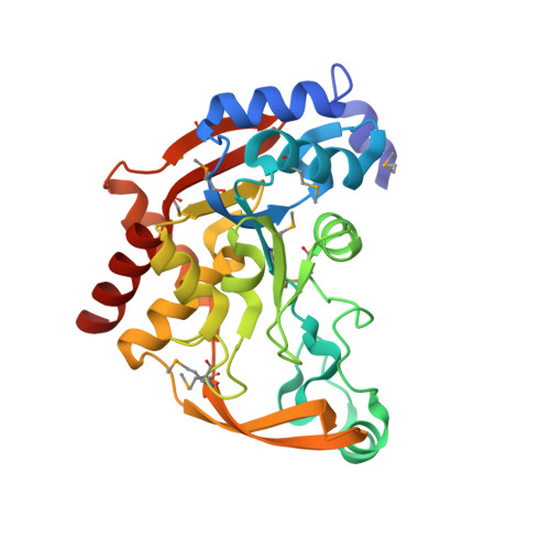 | |
UniProt | |||||
Entity Groups | |||||
| Sequence Clusters | 30% Identity50% Identity70% Identity90% Identity95% Identity100% Identity | ||||
| UniProt Group | A0A3P1UCA6 | ||||
Sequence AnnotationsExpand | |||||
Reference Sequence | |||||
| Ligands 3 Unique | |||||
|---|---|---|---|---|---|
| ID | Chains | Name / Formula / InChI Key | 2D Diagram | 3D Interactions | |
| ACO Download:Ideal Coordinates CCD File | F [auth A], H [auth B] | ACETYL COENZYME *A C23 H38 N7 O17 P3 S ZSLZBFCDCINBPY-ZSJPKINUSA-N |  | ||
| URA Download:Ideal Coordinates CCD File | G [auth A], I [auth B] | URACIL C4 H4 N2 O2 ISAKRJDGNUQOIC-UHFFFAOYSA-N |  | ||
| CL Download:Ideal Coordinates CCD File | C [auth A], D [auth A], E [auth A] | CHLORIDE ION Cl VEXZGXHMUGYJMC-UHFFFAOYSA-M |  | ||
| Modified Residues 1 Unique | |||||
|---|---|---|---|---|---|
| ID | Chains | Type | Formula | 2D Diagram | Parent |
| MSE Query on MSE | A, B | L-PEPTIDE LINKING | C5 H11 N O2 Se |  | MET |
| Length ( Å ) | Angle ( ˚ ) |
|---|---|
| a = 36.716 | α = 90 |
| b = 109.996 | β = 90 |
| c = 133.072 | γ = 90 |
| Software Name | Purpose |
|---|---|
| MD2 | data collection |
| HKL-3000 | phasing |
| MOLREP | phasing |
| REFMAC | refinement |
| Coot | model building |
| HKL-3000 | data reduction |
| HKL-3000 | data scaling |


