3SIO | pdb_00003sio

Ac-AChBP ligand binding domain (not including beta 9-10 linker) mutated to human alpha-7 nAChR


Experimental Data Snapshot

  • Method: X-RAY DIFFRACTION
  • Resolution: 2.32 Å
  • R-Value Free: 
    0.230 (Depositor), 0.224 (DCC) 
  • R-Value Work: 
    0.182 (Depositor), 0.178 (DCC) 
  • R-Value Observed: 
    0.184 (Depositor) 

Starting Model: in silico
View more details

wwPDB Validation 3D Report Full Report

Validation slider image for 3SIO

Ligand Structure Quality Assessment 


This is version 2.2 of the entry. See complete history

Literature

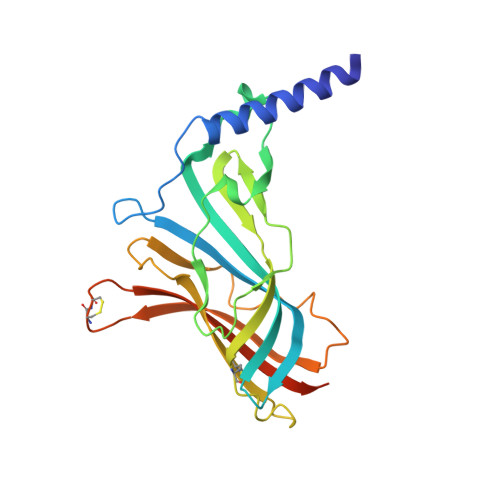
Creating an alpha-7 nicotinic acetylcholine recognition domain from the acetylcholine binding protein: crystallographic and ligand selectivity analyses

Nemecz, A.Taylor, P.

(2011) J Biological Chem 286: 42555-42565

  • DOI: https://doi.org/10.1074/jbc.M111.286583
  • Primary Citation Related Structures: 
    3SH1, 3SIO, 3T4M

  • PubMed Abstract: 

    Determining the structure of the ligand-binding domain of the nicotinic acetylcholine receptor (nAChR) has been a long standing goal in the design of selective drugs useful in implicated diseases for this prevalent receptor family. Acetylcholine-binding proteins have proven to be valuable surrogates with structural similarity and sequence identity to the extracellular domain of the nicotinic receptor, yet these soluble proteins have their unique features and do not serve as exact replicates of the nAChRs of interest. Here we systematically modify the sequence of these proteins toward the homomeric human α7 nAChR. These chimeric proteins exhibit a shift in affinities to reflect α7 binding characteristics yet maintain expression levels and stability conducive for crystallization. We also present a pentameric humanoid nAChR extracellular domain with the structural determination of the α7 nAChR glycosylation site.


  • Organizational Affiliation
    • Departments of Chemistry and Biochemistry, University of California, San Diego, La Jolla, California 92093-0650; Department of Pharmacology, Skaggs School of Pharmacy and Pharmaceutical Sciences, University of California, San Diego, La Jolla, California 92093-0650.

Macromolecule Content 

  • Total Structure Weight: 277.71 kDa 
  • Atom Count: 19,701 
  • Modeled Residue Count: 2,162 
  • Deposited Residue Count: 2,300 
  • Unique protein chains: 1

Macromolecules

Find similar proteins by:|  3D Structure
Entity ID: 1
MoleculeChains  Sequence LengthOrganismDetailsImage
Soluble acetylcholine receptor
A, B, C, D, E
A, B, C, D, E, F, G, H, I, J
230Aplysia californicaMutation(s): 18 
Gene Names: AChBP
UniProt
Find proteins for Q8WSF8 (Aplysia californica)
Explore Q8WSF8 
Go to UniProtKB:  Q8WSF8
Entity Groups
Sequence Clusters30% Identity50% Identity70% Identity90% Identity95% Identity100% Identity
UniProt GroupQ8WSF8
Glycosylation
Glycosylation Sites: 2
Sequence Annotations
Expand
Reference Sequence

Oligosaccharides

Help  
Entity ID: 2
MoleculeChains Length2D Diagram GlycosylationD Interactions
2-acetamido-2-deoxy-beta-D-glucopyranose-(1-4)-2-acetamido-2-deoxy-beta-D-glucopyranose
K, M, O
2N-Glycosylation
Glycosylation Resources
GlyTouCan: G42666HT
GlyCosmos: G42666HT
GlyGen: G42666HT
Entity ID: 3
MoleculeChains Length2D Diagram GlycosylationD Interactions
alpha-D-mannopyranose-(1-3)-[alpha-D-mannopyranose-(1-6)]alpha-D-mannopyranose-(1-6)-[alpha-D-mannopyranose-(1-3)]beta-D-mannopyranose-(1-4)-2-acetamido-2-deoxy-beta-D-glucopyranose-(1-4)-2-acetamido-2-deoxy-beta-D-glucopyranose
L, P
7N-Glycosylation
Glycosylation Resources
GlyTouCan: G55220VL
GlyCosmos: G55220VL
GlyGen: G55220VL
Entity ID: 4
MoleculeChains Length2D Diagram GlycosylationD Interactions
alpha-D-mannopyranose-(1-3)-alpha-D-mannopyranose-(1-6)-[alpha-D-mannopyranose-(1-3)]beta-D-mannopyranose-(1-4)-2-acetamido-2-deoxy-beta-D-glucopyranose-(1-4)-2-acetamido-2-deoxy-beta-D-glucopyranose
N, Q
6N-Glycosylation
Glycosylation Resources
GlyTouCan: G09724ZC
GlyCosmos: G09724ZC
GlyGen: G09724ZC

Small Molecules

Ligands 4 Unique
IDChains Name / Formula / InChI Key2D Diagram3D Interactions
MLK

Query on MLK


Download:Ideal Coordinates CCD File
AA [auth D]
DA [auth E]
FA [auth F]
HA [auth G]
KA [auth H]
AA [auth D],
DA [auth E],
FA [auth F],
HA [auth G],
KA [auth H],
NA [auth I],
QA [auth J],
S [auth A],
U [auth B],
X [auth C]
METHYLLYCACONITINE
C37 H50 N2 O10
XLTANAWLDBYGFU-VTLKBQQISA-N
NAG

Query on NAG


Download:Ideal Coordinates CCD File
CA [auth E]
EA [auth F]
IA [auth H]
LA [auth I]
PA [auth J]
CA [auth E],
EA [auth F],
IA [auth H],
LA [auth I],
PA [auth J],
R [auth A],
V [auth C]
2-acetamido-2-deoxy-beta-D-glucopyranose
C8 H15 N O6
OVRNDRQMDRJTHS-FMDGEEDCSA-N
MRD

Query on MRD


Download:Ideal Coordinates CCD File
BA [auth D]
GA [auth G]
JA [auth H]
OA [auth I]
T [auth B]
BA [auth D],
GA [auth G],
JA [auth H],
OA [auth I],
T [auth B],
W [auth C],
Z [auth D]
(4R)-2-METHYLPENTANE-2,4-DIOL
C6 H14 O2
SVTBMSDMJJWYQN-RXMQYKEDSA-N
MPD

Query on MPD


Download:Ideal Coordinates CCD File
MA [auth I],
Y [auth C]
(4S)-2-METHYL-2,4-PENTANEDIOL
C6 H14 O2
SVTBMSDMJJWYQN-YFKPBYRVSA-N

Experimental Data & Validation

Experimental Data

  • Method: X-RAY DIFFRACTION
  • Resolution: 2.32 Å
  • R-Value Free:  0.230 (Depositor), 0.224 (DCC) 
  • R-Value Work:  0.182 (Depositor), 0.178 (DCC) 
  • R-Value Observed: 0.184 (Depositor) 
Space Group: C 1 2 1
Unit Cell:
Length ( Å )Angle ( ˚ )
a = 143.218α = 90
b = 142.366β = 90.02
c = 144.477γ = 90
Software Package:
Software NamePurpose
CCP4model building
PHENIXrefinement
HKL-2000data reduction
HKL-2000data scaling
CCP4phasing

Structure Validation

View Full Validation Report


Ligand Structure Quality Assessment 


Entry History 

Deposition Data

Revision History  (Full details and data files)

  • Version 1.0: 2011-10-26
    Type: Initial release
  • Version 1.1: 2013-06-26
    Changes: Database references
  • Version 1.2: 2017-11-08
    Changes: Refinement description
  • Version 2.0: 2020-07-29
    Type: Remediation
    Reason: Carbohydrate remediation
    Changes: Atomic model, Data collection, Database references, Derived calculations, Structure summary
  • Version 2.1: 2024-04-03
    Changes: Data collection, Database references, Refinement description, Structure summary
  • Version 2.2: 2024-11-06
    Changes: Structure summary