


Crystal structure of putative acetyltransferase from Sphaerobacter thermophilus DSM 20745
Chang, C., Li, H., Clancy, S., Joachimiak, A.To be published.
Experimental Data Snapshot
Entity ID: 1 | |||||
|---|---|---|---|---|---|
| Molecule | Chains | Sequence Length | Organism | Details | Image |
| Putative acetyltransferase Sthe_0691 | 176 | Sphaerobacter thermophilus DSM 20745 | Mutation(s): 0 Gene Names: Sthe_0691 | 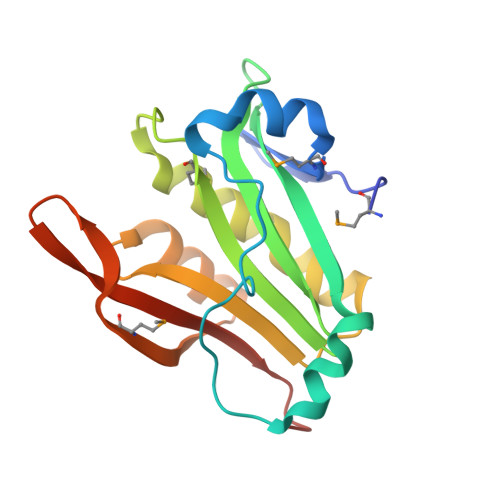 | |
UniProt | |||||
Entity Groups | |||||
| Sequence Clusters | 30% Identity50% Identity70% Identity90% Identity95% Identity100% Identity | ||||
| UniProt Group | D1C1L1 | ||||
Sequence AnnotationsExpand | |||||
Reference Sequence | |||||
| Ligands 1 Unique | |||||
|---|---|---|---|---|---|
| ID | Chains | Name / Formula / InChI Key | 2D Diagram | 3D Interactions | |
| SRT Download:Ideal Coordinates CCD File | C [auth B] | S,R MESO-TARTARIC ACID C4 H6 O6 FEWJPZIEWOKRBE-XIXRPRMCSA-N |  | ||
| Modified Residues 1 Unique | |||||
|---|---|---|---|---|---|
| ID | Chains | Type | Formula | 2D Diagram | Parent |
| MSE Query on MSE | A, B | L-PEPTIDE LINKING | C5 H11 N O2 Se |  | MET |
| Length ( Å ) | Angle ( ˚ ) |
|---|---|
| a = 89.196 | α = 90 |
| b = 89.196 | β = 90 |
| c = 115.726 | γ = 90 |
| Software Name | Purpose |
|---|---|
| REFMAC | refinement |
| PDB_EXTRACT | data extraction |
| SBC-Collect | data collection |
| HKL-3000 | data reduction |
| HKL-3000 | data scaling |
| HKL-3000 | phasing |
| MLPHARE | phasing |
| DM | phasing |
| SHELXDE | phasing |
| RESOLVE | phasing |
| ARP/wARP | model building |
| Coot | model building |


