3SEN | pdb_00003sen

Structure of Caskin1 Tandem SAMs


Experimental Data Snapshot

  • Method: X-RAY DIFFRACTION
  • Resolution: 3.10 Å
  • R-Value Free: 
    0.281 (Depositor), 0.259 (DCC) 
  • R-Value Work: 
    0.240 (Depositor), 0.237 (DCC) 
  • R-Value Observed: 
    0.242 (Depositor) 

Starting Model: experimental
View more details

wwPDB Validation 3D Report Full Report

Validation slider image for 3SEN

This is version 1.2 of the entry. See complete history

Literature

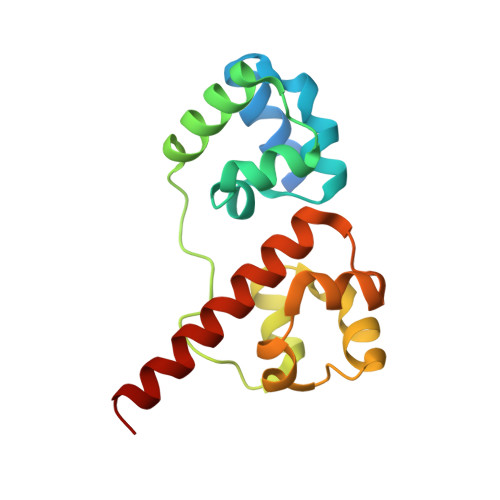
Tandem SAM domain structure of human Caskin1: a presynaptic, self-assembling scaffold for CASK.

Stafford, R.L.Hinde, E.Knight, M.J.Pennella, M.A.Ear, J.Digman, M.A.Gratton, E.Bowie, J.U.

(2011) Structure 19: 1826-1836

  • DOI: https://doi.org/10.1016/j.str.2011.09.018
  • Primary Citation Related Structures: 
    3SEI, 3SEN

  • PubMed Abstract: 

    The synaptic scaffolding proteins CASK and Caskin1 are part of the fibrous mesh of proteins that organize the active zones of neural synapses. CASK binds to a region of Caskin1 called the CASK interaction domain (CID). Adjacent to the CID, Caskin1 contains two tandem sterile α motif (SAM) domains. Many SAM domains form polymers so they are good candidates for forming the fibrous structures seen in the active zone. We show here that the SAM domains of Caskin1 form a new type of SAM helical polymer. The Caskin1 polymer interface exhibits a remarkable segregation of charged residues, resulting in a high sensitivity to ionic strength in vitro. The Caskin1 polymers can be decorated with CASK proteins, illustrating how these proteins may work together to organize the cytomatrix in active zones.


  • Organizational Affiliation
    • Department of Chemistry and Biochemistry, UCLA-DOE Institute of Genomics and Proteomics, Molecular Biology Institute, University of California, Los Angeles, Los Angeles, CA 90095-1570, USA.

Macromolecule Content 

  • Total Structure Weight: 71.61 kDa 
  • Atom Count: 4,392 
  • Modeled Residue Count: 554 
  • Deposited Residue Count: 628 
  • Unique protein chains: 1

Macromolecules

Find similar proteins by:|  3D Structure
Entity ID: 1
MoleculeChains  Sequence LengthOrganismDetailsImage
Caskin-1
A, B, C, D
157Homo sapiensMutation(s): 0 
Gene Names: CASKIN1KIAA1306
UniProt & NIH Common Fund Data Resources
Find proteins for Q8WXD9 (Homo sapiens)
Explore Q8WXD9 
Go to UniProtKB:  Q8WXD9
PHAROS:  Q8WXD9
GTEx:  ENSG00000167971 
Entity Groups
Sequence Clusters30% Identity50% Identity70% Identity90% Identity95% Identity100% Identity
UniProt GroupQ8WXD9
Sequence Annotations
Expand
Reference Sequence

Small Molecules

Ligands 1 Unique
IDChains Name / Formula / InChI Key2D Diagram3D Interactions
K

Query on K


Download:Ideal Coordinates CCD File
E [auth A]POTASSIUM ION
K
NPYPAHLBTDXSSS-UHFFFAOYSA-N

Experimental Data & Validation

Experimental Data

  • Method: X-RAY DIFFRACTION
  • Resolution: 3.10 Å
  • R-Value Free:  0.281 (Depositor), 0.259 (DCC) 
  • R-Value Work:  0.240 (Depositor), 0.237 (DCC) 
  • R-Value Observed: 0.242 (Depositor) 
Space Group: P 21 21 21
Unit Cell:
Length ( Å )Angle ( ˚ )
a = 92.226α = 90
b = 94.597β = 90
c = 118.633γ = 90
Software Package:
Software NamePurpose
SCALEPACKdata scaling
REFMACrefinement
PDB_EXTRACTdata extraction
ADSCdata collection
DENZOdata reduction
PHASERphasing

Structure Validation

View Full Validation Report


Entry History 

Deposition Data

Revision History  (Full details and data files)

  • Version 1.0: 2011-12-14
    Type: Initial release
  • Version 1.1: 2012-03-28
    Changes: Database references
  • Version 1.2: 2023-09-13
    Changes: Data collection, Database references, Refinement description