3SDE

Crystal structure of a paraspeckle-protein heterodimer, PSPC1/NONO


Experimental Data Snapshot

  • Method: X-RAY DIFFRACTION
  • Resolution: 1.90 Å
  • R-Value Free: 0.228 
  • R-Value Work: 0.183 
  • R-Value Observed: 0.185 

wwPDB Validation   3D Report Full Report


This is version 1.3 of the entry. See complete history


Literature

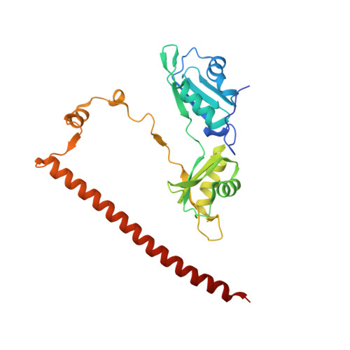
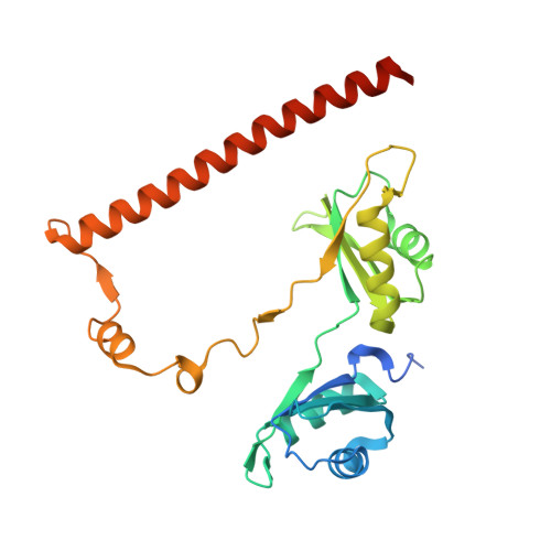
Structure of the heterodimer of human NONO and paraspeckle protein component 1 and analysis of its role in subnuclear body formation.

Passon, D.M.Lee, M.Rackham, O.Stanley, W.A.Sadowska, A.Filipovska, A.Fox, A.H.Bond, C.S.

(2012) Proc Natl Acad Sci U S A 109: 4846-4850

  • DOI: https://doi.org/10.1073/pnas.1120792109
  • Primary Citation of Related Structures:  
    3SDE

  • PubMed Abstract: 

    Proteins of the Drosophila behavior/human splicing (DBHS) family include mammalian SFPQ (PSF), NONO (p54nrb), PSPC1, and invertebrate NONA and Hrp65. DBHS proteins are predominately nuclear, and are involved in transcriptional and posttranscriptional gene regulatory functions as well as DNA repair. DBHS proteins influence a wide gamut of biological processes, including the regulation of circadian rhythm, carcinogenesis, and progression of cancer. Additionally, mammalian DBHS proteins associate with the architectural long noncoding RNA NEAT1 (Menε/β) to form paraspeckles, subnuclear bodies that alter gene expression via the nuclear retention of RNA. Here we describe the crystal structure of the heterodimer of the multidomain conserved region of the DBHS proteins, PSPC1 and NONO. These proteins form an extensively intertwined dimer, consistent with the observation that the different DBHS proteins are typically copurified from mammalian cells, and suggesting that they act as obligate heterodimers. The PSPC1/NONO heterodimer has a right-handed antiparallel coiled-coil that positions two of four RNA recognition motif domains in an unprecedented arrangement on either side of a 20-Å channel. This configuration is supported by a protein:protein interaction involving the NONA/paraspeckle domain, which is characteristic of the DBHS family. By examining various mutants and truncations in cell culture, we find that DBHS proteins require an additional antiparallel coiled-coil emanating from either end of the dimer for paraspeckle subnuclear body formation. These results suggest that paraspeckles may potentially form through self-association of DBHS dimers into higher-order structures.


  • Organizational Affiliation

    School of Chemistry and Biochemistry, University of Western Australia, Crawley, Western Australia 6009, Australia.


Macromolecules
Find similar proteins by:  (by identity cutoff)  |  3D Structure
Entity ID: 1
MoleculeChains Sequence LengthOrganismDetailsImage
Paraspeckle component 1261Homo sapiensMutation(s): 0 
Gene Names: PSPC1PSP1
UniProt & NIH Common Fund Data Resources
Find proteins for Q8WXF1 (Homo sapiens)
Explore Q8WXF1 
Go to UniProtKB:  Q8WXF1
PHAROS:  Q8WXF1
GTEx:  ENSG00000121390 
Entity Groups  
Sequence Clusters30% Identity50% Identity70% Identity90% Identity95% Identity100% Identity
UniProt GroupQ8WXF1
Sequence Annotations
Expand
  • Reference Sequence
Find similar proteins by:  (by identity cutoff)  |  3D Structure
Entity ID: 2
MoleculeChains Sequence LengthOrganismDetailsImage
Non-POU domain-containing octamer-binding protein261Homo sapiensMutation(s): 0 
Gene Names: NONONRB54
UniProt & NIH Common Fund Data Resources
Find proteins for Q15233 (Homo sapiens)
Explore Q15233 
Go to UniProtKB:  Q15233
PHAROS:  Q15233
GTEx:  ENSG00000147140 
Entity Groups  
Sequence Clusters30% Identity50% Identity70% Identity90% Identity95% Identity100% Identity
UniProt GroupQ15233
Sequence Annotations
Expand
  • Reference Sequence
Small Molecules
Ligands 1 Unique
IDChains Name / Formula / InChI Key2D Diagram3D Interactions
EDO
Query on EDO

Download Ideal Coordinates CCD File 
C [auth A]
D [auth A]
E [auth A]
F [auth A]
G [auth A]
C [auth A],
D [auth A],
E [auth A],
F [auth A],
G [auth A],
H [auth B],
I [auth B],
J [auth B],
K [auth B],
L [auth B],
M [auth B]
1,2-ETHANEDIOL
C2 H6 O2
LYCAIKOWRPUZTN-UHFFFAOYSA-N
Experimental Data & Validation

Experimental Data

  • Method: X-RAY DIFFRACTION
  • Resolution: 1.90 Å
  • R-Value Free: 0.228 
  • R-Value Work: 0.183 
  • R-Value Observed: 0.185 
  • Space Group: C 1 2 1
Unit Cell:
Length ( Å )Angle ( ˚ )
a = 90.9α = 90
b = 67.18β = 99.96
c = 94.08γ = 90
Software Package:
Software NamePurpose
ADSCdata collection
SOLVEphasing
BUSTERrefinement
XDSdata reduction
SCALAdata scaling

Structure Validation

View Full Validation Report


Entry History 

Deposition Data

Revision History  (Full details and data files)

  • Version 1.0: 2012-03-14
    Type: Initial release
  • Version 1.1: 2012-03-28
    Changes: Database references
  • Version 1.2: 2012-04-11
    Changes: Database references
  • Version 1.3: 2024-02-28
    Changes: Data collection, Database references, Derived calculations