3SCH | pdb_00003sch

Co(II)-HppE with R-HPP


Experimental Data Snapshot

  • Method: X-RAY DIFFRACTION
  • Resolution: 2.10 Å
  • R-Value Free: 
    0.245 (Depositor), 0.247 (DCC) 
  • R-Value Work: 
    0.217 (Depositor), 0.222 (DCC) 
  • R-Value Observed: 
    0.217 (Depositor) 

Starting Model: experimental
View more details

wwPDB Validation 3D Report Full Report

Validation slider image for 3SCH

This is version 1.6 of the entry. See complete history

Literature

Structural basis of regiospecificity of a mononuclear iron enzyme in antibiotic fosfomycin biosynthesis.

Yun, D.Dey, M.Higgins, L.J.Yan, F.Liu, H.W.Drennan, C.L.

(2011) J Am Chem Soc 133: 11262-11269

  • DOI: https://doi.org/10.1021/ja2025728
  • Primary Citation Related Structures: 
    3SCF, 3SCG, 3SCH

  • PubMed Abstract: 

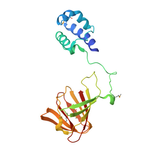
    Hydroxypropylphosphonic acid epoxidase (HppE) is an unusual mononuclear iron enzyme that uses dioxygen to catalyze the oxidative epoxidation of (S)-2-hydroxypropylphosphonic acid (S-HPP) in the biosynthesis of the antibiotic fosfomycin. Additionally, the enzyme converts the R-enantiomer of the substrate (R-HPP) to 2-oxo-propylphosphonic acid. To probe the mechanism of HppE regiospecificity, we determined three X-ray structures: R-HPP with inert cobalt-containing enzyme (Co(II)-HppE) at 2.1 Å resolution; R-HPP with active iron-containing enzyme (Fe(II)-HppE) at 3.0 Å resolution; and S-HPP-Fe(II)-HppE in complex with dioxygen mimic NO at 2.9 Å resolution. These structures, along with previously determined structures of S-HPP-HppE, identify the dioxygen binding site on iron and elegantly illustrate how HppE is able to recognize both substrate enantiomers to catalyze two completely distinct reactions.


  • Organizational Affiliation
    • Department of Chemistry, Massachusetts Institute of Technology, Cambridge, Massachusetts 02139, USA.

Macromolecule Content 

  • Total Structure Weight: 43.59 kDa 
  • Atom Count: 3,069 
  • Modeled Residue Count: 387 
  • Deposited Residue Count: 396 
  • Unique protein chains: 1

Macromolecules

Find similar proteins by:|  3D Structure
Entity ID: 1
MoleculeChains  Sequence LengthOrganismDetailsImage
Epoxidase
A, B
198Streptomyces wedmorensisMutation(s): 0 
Gene Names: fom4
EC: 1.11.1.23
UniProt
Find proteins for Q56185 (Streptomyces wedmorensis)
Explore Q56185 
Go to UniProtKB:  Q56185
Entity Groups
Sequence Clusters30% Identity50% Identity70% Identity90% Identity95% Identity100% Identity
UniProt GroupQ56185
Sequence Annotations
Expand
Reference Sequence

Experimental Data & Validation

Experimental Data

  • Method: X-RAY DIFFRACTION
  • Resolution: 2.10 Å
  • R-Value Free:  0.245 (Depositor), 0.247 (DCC) 
  • R-Value Work:  0.217 (Depositor), 0.222 (DCC) 
  • R-Value Observed: 0.217 (Depositor) 
Space Group: P 65 2 2
Unit Cell:
Length ( Å )Angle ( ˚ )
a = 86.12α = 90
b = 86.12β = 90
c = 219.056γ = 120
Software Package:
Software NamePurpose
ADSCdata collection
CNSrefinement
DENZOdata reduction
SCALEPACKdata scaling
CNSphasing

Structure Validation

View Full Validation Report


Entry History 

Deposition Data

Revision History  (Full details and data files)

  • Version 1.0: 2011-07-06
    Type: Initial release
  • Version 1.1: 2011-07-13
    Changes: Version format compliance
  • Version 1.2: 2011-08-10
    Changes: Database references
  • Version 1.3: 2019-11-20
    Changes: Derived calculations
  • Version 1.4: 2023-09-13
    Changes: Data collection, Database references, Derived calculations, Refinement description
  • Version 1.5: 2023-12-06
    Changes: Data collection
  • Version 1.6: 2024-11-20
    Changes: Structure summary