3S9C | pdb_00003s9c

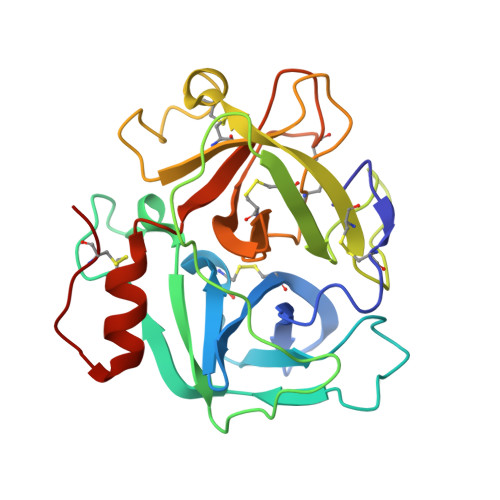
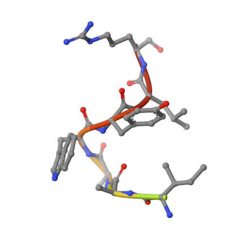
Russell's viper venom serine proteinase, RVV-V in complex with the fragment (residues 1533-1546) of human factor V


Experimental Data Snapshot

  • Method: X-RAY DIFFRACTION
  • Resolution: 1.80 Å
  • R-Value Free: 
    0.218 (Depositor), 0.210 (DCC) 
  • R-Value Work: 
    0.183 (Depositor), 0.180 (DCC) 
  • R-Value Observed: 
    0.183 (Depositor) 

Starting Model: experimental
View more details

wwPDB Validation   3D Report Full Report


Ligand Structure Quality Assessment 

Created with Raphaël 2.3.0Worse 01 BetterLigand structure goodness of fit to experimental dataBest fitted GLCClick on this verticalbar to view detailsBest fitted BGCClick on this verticalbar to view details

This is version 2.2 of the entry. See complete history


Literature

Structural basis of coagulation factor V recognition for cleavage by RVV-V

Nakayama, D.Ben Ammar, Y.Miyata, T.Takeda, S.

(2011) FEBS Lett 585: 3020-3025

  • DOI: https://doi.org/10.1016/j.febslet.2011.08.022
  • Primary Citation of Related Structures:  
    3S9A, 3S9B, 3S9C, 3SBK

  • PubMed Abstract: 

    Russell's viper venom factor V (FV) activator (RVV-V) is a thrombin-like proteinase that specifically cleaves the Arg1545-Ser1546 bond of FV. Here we present the crystal structure of RVV-V in complex with the FV14 peptide (residues 1533-1546 of human FV) determined at 1.8Å resolution. The structure reveals multiple interactions between RVV-V and the seven residues, Ile1539 (P(7))-Arg1545 (P(1)), of the cleaved substrate. Comparison with substrate-free structures reveals conformational changes of the RVV-V loops upon substrate binding, suggesting that the multiple interactions are mediated by an induced-fit mechanism. The results provide an explanation for the narrow specificity of RVV-V.


  • Organizational Affiliation

    Department of Cardiac Physiology, National Cerebral and Cardiovascular Center Research Institute, Suita, Osaka, Japan.


Macromolecules
Find similar proteins by:  (by identity cutoff)  |  3D Structure
Entity ID: 1
MoleculeChains Sequence LengthOrganismDetailsImage
Vipera russelli proteinase RVV-V gamma234Daboia siamensisMutation(s): 0 
EC: 3.4.21.95
UniProt
Find proteins for P18965 (Daboia siamensis)
Explore P18965 
Go to UniProtKB:  P18965
Entity Groups  
Sequence Clusters30% Identity50% Identity70% Identity90% Identity95% Identity100% Identity
UniProt GroupP18965
Glycosylation
Glycosylation Sites: 1
Sequence Annotations
Expand
  • Reference Sequence

Find similar proteins by:  Sequence   |   3D Structure  

Entity ID: 2
MoleculeChains Sequence LengthOrganismDetailsImage
Coagulation factor V14Homo sapiensMutation(s): 0 
UniProt & NIH Common Fund Data Resources
Find proteins for P12259 (Homo sapiens)
Explore P12259 
Go to UniProtKB:  P12259
PHAROS:  P12259
GTEx:  ENSG00000198734 
Entity Groups  
Sequence Clusters30% Identity50% Identity70% Identity90% Identity95% Identity100% Identity
UniProt GroupP12259
Sequence Annotations
Expand
  • Reference Sequence
Oligosaccharides

Help

Entity ID: 3
MoleculeChains Length2D Diagram Glycosylation3D Interactions
beta-D-mannopyranose-(1-4)-2-acetamido-2-deoxy-beta-D-glucopyranose-(1-4)-2-acetamido-2-deoxy-beta-D-glucopyranose
C
3N-Glycosylation
Glycosylation Resources
GlyTouCan:  G15407YE
GlyCosmos:  G15407YE
GlyGen:  G15407YE
Small Molecules
Ligands 4 Unique
IDChains Name / Formula / InChI Key2D Diagram3D Interactions
GLC
Query on GLC

Download Ideal Coordinates CCD File 
E [auth A]alpha-D-glucopyranose
C6 H12 O6
WQZGKKKJIJFFOK-DVKNGEFBSA-N
BGC
Query on BGC

Download Ideal Coordinates CCD File 
D [auth A]beta-D-glucopyranose
C6 H12 O6
WQZGKKKJIJFFOK-VFUOTHLCSA-N
ZN
Query on ZN

Download Ideal Coordinates CCD File 
J [auth A],
K [auth A],
L [auth A]
ZINC ION
Zn
PTFCDOFLOPIGGS-UHFFFAOYSA-N
ACT
Query on ACT

Download Ideal Coordinates CCD File 
F [auth A],
G [auth A],
H [auth A],
I [auth A]
ACETATE ION
C2 H3 O2
QTBSBXVTEAMEQO-UHFFFAOYSA-M
Experimental Data & Validation

Experimental Data

  • Method: X-RAY DIFFRACTION
  • Resolution: 1.80 Å
  • R-Value Free:  0.218 (Depositor), 0.210 (DCC) 
  • R-Value Work:  0.183 (Depositor), 0.180 (DCC) 
  • R-Value Observed: 0.183 (Depositor) 
Space Group: P 61
Unit Cell:
Length ( Å )Angle ( ˚ )
a = 101.2α = 90
b = 101.2β = 90
c = 44.2γ = 120
Software Package:
Software NamePurpose
CNSrefinement
HKL-2000data collection
HKL-2000data reduction
HKL-2000data scaling
MOLREPphasing

Structure Validation

View Full Validation Report


Ligand Structure Quality Assessment 

Created with Raphaël 2.3.0Worse 01 BetterLigand structure goodness of fit to experimental dataBest fitted GLCClick on this verticalbar to view detailsBest fitted BGCClick on this verticalbar to view details

Entry History 

Deposition Data

Revision History  (Full details and data files)

  • Version 1.0: 2011-09-07
    Type: Initial release
  • Version 1.1: 2013-07-03
    Changes: Database references
  • Version 2.0: 2020-07-29
    Type: Remediation
    Reason: Carbohydrate remediation
    Changes: Atomic model, Data collection, Database references, Derived calculations, Structure summary
  • Version 2.1: 2023-11-01
    Changes: Data collection, Database references, Refinement description, Structure summary
  • Version 2.2: 2024-10-09
    Changes: Structure summary