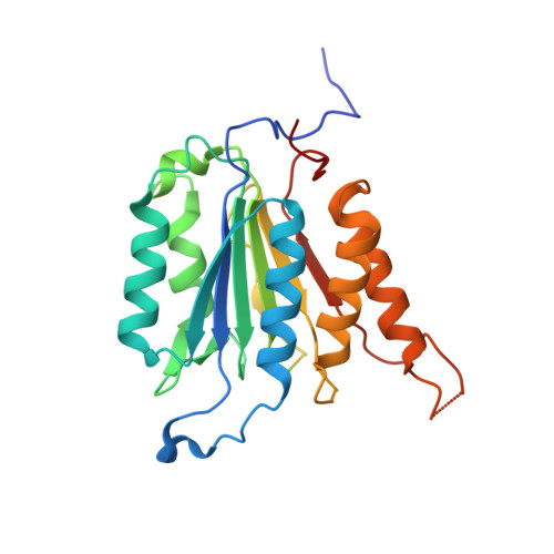
3S8E

Phosphorylation regulates assembly of the caspase-6 substrate-binding groove


Experimental Data Snapshot

  • Method: X-RAY DIFFRACTION
  • Resolution: 2.88 Å
  • R-Value Free: 0.256 
  • R-Value Work: 0.216 
  • R-Value Observed: 0.218 

Starting Model: experimental
View more details

wwPDB Validation   3D Report Full Report


This is version 1.2 of the entry. See complete history


Literature

Phosphorylation regulates assembly of the caspase-6 substrate-binding groove.

Velazquez-Delgado, E.M.Hardy, J.A.

(2012) Structure 20: 742-751

  • DOI: https://doi.org/10.1016/j.str.2012.02.003
  • Primary Citation of Related Structures:  
    3S8E

  • PubMed Abstract: 

    Caspases, a family of apoptotic proteases, are increasingly recognized as being extensively phosphorylated, usually leading to inactivation. To date, no structural mechanism for phosphorylation-based caspase inactivation is available, although this information may be key to achieving caspase-specific inhibition. Caspase-6 has recently been implicated in neurodegenerative conditions including Huntington's and Alzheimer's diseases. A full understanding of caspase-6 regulation is crucial to caspase-6-specific inhibition. Caspase-6 is phosphorylated by ARK5 kinase at serine 257 leading to suppression of cell death via caspase-6 inhibition. Our structure of the fully inactive phosphomimetic S257D reveals that phosphorylation results in a steric clash with P201 in the L2' loop. Removal of the proline side chain alleviates the clash resulting in nearly wild-type activity levels. This phosphomimetic-mediated steric clash causes misalignment of the substrate-binding groove, preventing substrate binding. Substrate-binding loop misalignment appears to be a widely used regulatory strategy among caspases and may present a new paradigm for caspase-specific control.


  • Organizational Affiliation

    Department of Chemistry, University of Massachusetts Amherst, Amherst, MA 01003, USA.


Macromolecules
Find similar proteins by:  (by identity cutoff)  |  3D Structure
Entity ID: 1
MoleculeChains Sequence LengthOrganismDetailsImage
Caspase-6
A, B, C, D, E
A, B, C, D, E, F, G, H
277Homo sapiensMutation(s): 1 
Gene Names: CASP6MCH2
EC: 3.4.22.59
UniProt & NIH Common Fund Data Resources
Find proteins for P55212 (Homo sapiens)
Explore P55212 
Go to UniProtKB:  P55212
PHAROS:  P55212
GTEx:  ENSG00000138794 
Entity Groups  
Sequence Clusters30% Identity50% Identity70% Identity90% Identity95% Identity100% Identity
UniProt GroupP55212
Sequence Annotations
Expand
  • Reference Sequence
Experimental Data & Validation

Experimental Data

  • Method: X-RAY DIFFRACTION
  • Resolution: 2.88 Å
  • R-Value Free: 0.256 
  • R-Value Work: 0.216 
  • R-Value Observed: 0.218 
  • Space Group: P 1 21 1
Unit Cell:
Length ( Å )Angle ( ˚ )
a = 81.671α = 90
b = 163.662β = 94.17
c = 89.015γ = 90
Software Package:
Software NamePurpose
CrystalCleardata collection
PHASERphasing
REFMACrefinement
HKL-2000data reduction
SCALEPACKdata scaling

Structure Validation

View Full Validation Report


Entry History 

Deposition Data

Revision History  (Full details and data files)

  • Version 1.0: 2012-04-11
    Type: Initial release
  • Version 1.1: 2013-07-10
    Changes: Database references
  • Version 1.2: 2023-11-01
    Changes: Data collection, Database references, Refinement description