3S46 | pdb_00003s46

The crystal structure of alanine racemase from streptococcus pneumoniae


Experimental Data Snapshot

  • Method: X-RAY DIFFRACTION
  • Resolution: 2.00 Å
  • R-Value Free: 
    0.200 (Depositor), 0.194 (DCC) 
  • R-Value Work: 
    0.168 (Depositor), 0.164 (DCC) 
  • R-Value Observed: 
    0.169 (Depositor) 

Starting Model: experimental
View more details

wwPDB Validation 3D Report Full Report

Validation slider image for 3S46

This is version 1.4 of the entry. See complete history

Literature

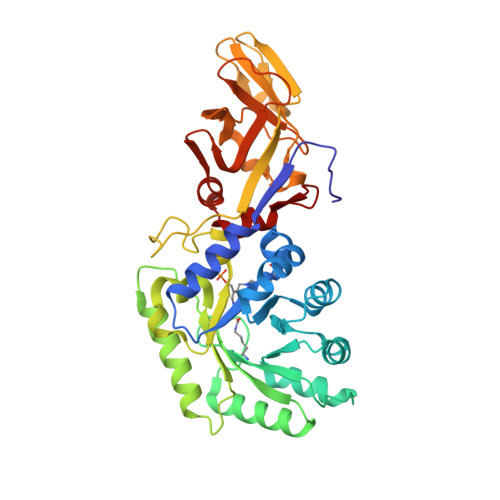
The crystal structure of alanine racemase from Streptococcus pneumoniae, a target for structure-based drug design.

Im, H.Sharpe, M.L.Strych, U.Davlieva, M.Krause, K.L.

(2011) BMC Microbiol 11: 116-116

  • DOI: https://doi.org/10.1186/1471-2180-11-116
  • Primary Citation Related Structures: 
    3S46

  • PubMed Abstract: 

    Streptococcus pneumoniae is a globally important pathogen. The Gram-positive diplococcus is a leading cause of pneumonia, otitis media, bacteremia, and meningitis, and antibiotic resistant strains have become increasingly common over recent years. Alanine racemase is a ubiquitous enzyme among bacteria and provides the essential cell wall precursor, D-alanine. Since it is absent in humans, this enzyme is an attractive target for the development of drugs against S. pneumoniae and other bacterial pathogens. Here we report the crystal structure of alanine racemase from S. pneumoniae (AlrSP). Crystals diffracted to a resolution of 2.0 Å and belong to the space group P3121 with the unit cell parameters a = b = 119.97 Å, c = 118.10 Å, α = β = 90° and γ = 120°. Structural comparisons show that AlrSP shares both an overall fold and key active site residues with other bacterial alanine racemases. The active site cavity is similar to other Gram positive alanine racemases, featuring a restricted but conserved entryway. We have solved the structure of AlrSP, an essential step towards the development of an accurate pharmacophore model of the enzyme, and an important contribution towards our on-going alanine racemase structure-based drug design project. We have identified three regions on the enzyme that could be targeted for inhibitor design, the active site, the dimer interface, and the active site entryway.


  • Organizational Affiliation
    • Research Institute of Pharmaceutical Sciences, College of Pharmacy, Seoul National University, Seoul, Korea.

Macromolecule Content 

  • Total Structure Weight: 80.46 kDa 
  • Atom Count: 6,161 
  • Modeled Residue Count: 734 
  • Deposited Residue Count: 734 
  • Unique protein chains: 1

Macromolecules

Find similar proteins by:|  3D Structure
Entity ID: 1
MoleculeChains  Sequence LengthOrganismDetailsImage
Alanine racemase
A, B
367Streptococcus pneumoniaeMutation(s): 0 
Gene Names: alaRalrSP_1698
EC: 5.1.1.1
UniProt
Find proteins for P0A2W8 (Streptococcus pneumoniae serotype 4 (strain ATCC BAA-334 / TIGR4))
Explore P0A2W8 
Go to UniProtKB:  P0A2W8
Entity Groups
Sequence Clusters30% Identity50% Identity70% Identity90% Identity95% Identity100% Identity
UniProt GroupP0A2W8
Sequence Annotations
Expand
Reference Sequence

Small Molecules

Ligands 1 Unique
IDChains Name / Formula / InChI Key2D Diagram3D Interactions
BEZ

Query on BEZ


Download:Ideal Coordinates CCD File
C [auth A]BENZOIC ACID
C7 H6 O2
WPYMKLBDIGXBTP-UHFFFAOYSA-N
Modified Residues  2 Unique
IDChains TypeFormula2D DiagramParent
KCX
Query on KCX
A, B
L-PEPTIDE LINKINGC7 H14 N2 O4LYS
LLP
Query on LLP
A, B
L-PEPTIDE LINKINGC14 H22 N3 O7 PLYS

Experimental Data & Validation

Experimental Data

  • Method: X-RAY DIFFRACTION
  • Resolution: 2.00 Å
  • R-Value Free:  0.200 (Depositor), 0.194 (DCC) 
  • R-Value Work:  0.168 (Depositor), 0.164 (DCC) 
  • R-Value Observed: 0.169 (Depositor) 
Space Group: P 31 2 1
Unit Cell:
Length ( Å )Angle ( ˚ )
a = 119.973α = 90
b = 119.973β = 90
c = 118.099γ = 120
Software Package:
Software NamePurpose
DENZOdata reduction
SCALEPACKdata scaling
CNSrefinement
REFMACrefinement
PDB_EXTRACTdata extraction
CNSphasing

Structure Validation

View Full Validation Report


Entry History 

Deposition Data

Revision History  (Full details and data files)

  • Version 1.0: 2011-06-22
    Type: Initial release
  • Version 1.1: 2011-07-13
    Changes: Version format compliance
  • Version 1.2: 2017-11-08
    Changes: Refinement description
  • Version 1.3: 2023-11-01
    Changes: Data collection, Database references, Derived calculations, Refinement description
  • Version 1.4: 2023-12-06
    Changes: Data collection