3S3G | pdb_00003s3g

Crystal Structure of Human Aldose Reductase Complexed with Tolmetin


Experimental Data Snapshot

  • Method: X-RAY DIFFRACTION
  • Resolution: 1.80 Å
  • R-Value Free: 
    0.204 (Depositor), 0.201 (DCC) 
  • R-Value Work: 
    0.160 (Depositor), 0.159 (DCC) 
  • R-Value Observed: 
    0.163 (Depositor) 

Starting Model: experimental
View more details

wwPDB Validation 3D Report Full Report

Validation slider image for 3S3G

Ligand Structure Quality Assessment 


This is version 1.1 of the entry. See complete history

Literature

The molecular basis for inhibition of sulindac and its metabolites towards human aldose reductase.

Zheng, X.Zhang, L.Zhai, J.Chen, Y.Luo, H.Hu, X.

(2012) FEBS Lett 586: 55-59

  • DOI: https://doi.org/10.1016/j.febslet.2011.11.023
  • Primary Citation Related Structures: 
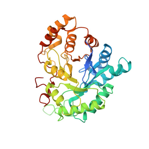
    3RX2, 3RX3, 3RX4, 3S3G

  • PubMed Abstract: 

    Sulindac (SLD) exhibits both the highest inhibitory activity towards human aldose reductase (AR) among popular non-steroidal anti-inflammatory drugs and clear beneficial clinical effects on Type 2 diabetes. However, the molecular basis for these properties is unclear. Here, we report that SLD and its pharmacologically active/inactive metabolites, SLD sulfide and SLD sulfone, are equally effective as un-competitive inhibitors of AR in vitro. Crystallographic analysis reveals that π-π stacking favored by the distinct scaffold of SLDs is pivotal to their high AR inhibitory activities. These results also suggest that SLD sulfone could be a potent lead compound for AR inhibition in vivo.


  • Organizational Affiliation
    • School of Pharmaceutical Sciences, Sun Yat-sen University, Guangzhou 510006, China. huxpeng@mail.sysu.edu.cn

Macromolecule Content 

  • Total Structure Weight: 39.07 kDa 
  • Atom Count: 3,055 
  • Modeled Residue Count: 316 
  • Deposited Residue Count: 336 
  • Unique protein chains: 1

Macromolecules

Find similar proteins by:|  3D Structure
Entity ID: 1
MoleculeChains  Sequence LengthOrganismDetailsImage
Aldose reductase336Homo sapiensMutation(s): 0 
Gene Names: AKR1B1ALDR1
EC: 1.1.1.21 (PDB Primary Data), 1.1.1.372 (UniProt), 1.1.1.300 (UniProt), 1.1.1.54 (UniProt)
UniProt & NIH Common Fund Data Resources
Find proteins for P15121 (Homo sapiens)
Explore P15121 
Go to UniProtKB:  P15121
PHAROS:  P15121
GTEx:  ENSG00000085662 
Entity Groups
Sequence Clusters30% Identity50% Identity70% Identity90% Identity95% Identity100% Identity
UniProt GroupP15121
Sequence Annotations
Expand
Reference Sequence

Small Molecules

Ligands 2 Unique
IDChains Name / Formula / InChI Key2D Diagram3D Interactions
NAP

Query on NAP


Download:Ideal Coordinates CCD File
B [auth A]NADP NICOTINAMIDE-ADENINE-DINUCLEOTIDE PHOSPHATE
C21 H28 N7 O17 P3
XJLXINKUBYWONI-NNYOXOHSSA-N
TLT

Query on TLT


Download:Ideal Coordinates CCD File
C [auth A]Tolmetin
C15 H15 N O3
UPSPUYADGBWSHF-UHFFFAOYSA-N
Binding Affinity Annotations 
IDSourceBinding Affinity
TLT BindingDB:  3S3G IC50: 2390 (nM) from 1 assay(s)

Experimental Data & Validation

Experimental Data

  • Method: X-RAY DIFFRACTION
  • Resolution: 1.80 Å
  • R-Value Free:  0.204 (Depositor), 0.201 (DCC) 
  • R-Value Work:  0.160 (Depositor), 0.159 (DCC) 
  • R-Value Observed: 0.163 (Depositor) 
Space Group: P 1 21 1
Unit Cell:
Length ( Å )Angle ( ˚ )
a = 47.132α = 90
b = 66.571β = 92.02
c = 49.29γ = 90
Software Package:
Software NamePurpose
CrysalisProdata collection
MOLREPphasing
PHENIXrefinement
CrysalisProdata reduction

Structure Validation

View Full Validation Report


Ligand Structure Quality Assessment 


Entry History 

Deposition Data

Revision History  (Full details and data files)

  • Version 1.0: 2012-04-04
    Type: Initial release
  • Version 1.1: 2023-11-01
    Changes: Data collection, Database references, Derived calculations, Refinement description