3S33

Structure of Thermus thermophilus cytochrome ba3 oxidase 10s after Xe depressurization


Experimental Data Snapshot

  • Method: X-RAY DIFFRACTION
  • Resolution: 4.45 Å
  • R-Value Free: 0.350 
  • R-Value Work: 0.305 
  • R-Value Observed: 0.307 

wwPDB Validation   3D Report Full Report


Ligand Structure Quality Assessment 


This is version 1.3 of the entry. See complete history


Literature

Mobility of Xe atoms within the oxygen diffusion channel of cytochrome ba(3) oxidase.

Luna, V.M.Fee, J.A.Deniz, A.A.Stout, C.D.

(2012) Biochemistry 51: 4669-4676

  • DOI: https://doi.org/10.1021/bi3003988
  • Primary Citation of Related Structures:  
    3S33, 3S38, 3S39, 3S3A, 3S3B, 3S3C, 3S3D

  • PubMed Abstract: 

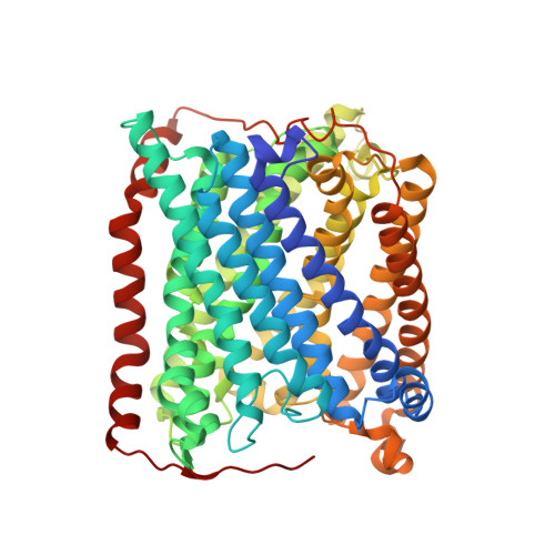
    We use a form of "freeze-trap, kinetic crystallography" to explore the migration of Xe atoms away from the dinuclear heme a(3)/Cu(B) center in Thermus thermophilus cytochrome ba(3) oxidase. This enzyme is a member of the heme-copper oxidase superfamily and is thus crucial for dioxygen-dependent life. The mechanisms involved in the migration of oxygen, water, electrons, and protons into and/or out of the specialized channels of the heme-copper oxidases are generally not well understood. Pressurization of crystals with Xe gas previously revealed a O(2) diffusion channel in cytochrome ba(3) oxidase that is continuous, Y-shaped, 18-20 Å in length and comprised of hydrophobic residues, connecting the protein surface within the bilayer to the a(3)-Cu(B) center in the active site. To understand movement of gas molecules within the O(2) channel, we performed crystallographic analysis of 19 Xe laden crystals freeze-trapped in liquid nitrogen at selected times between 0 and 480 s while undergoing outgassing at room temperature. Variation in Xe crystallographic occupancy at five discrete sites as a function of time leads to a kinetic model revealing relative degrees of mobility of Xe atoms within the channel. Xe egress occurs primarily through the channel formed by the Xe1 → Xe5 → Xe3 → Xe4 sites, suggesting that ingress of O(2) is likely to occur by the reverse of this process. The channel itself appears not to undergo significant structural changes during Xe migration, thereby indicating a passive role in this important physiological function.


  • Organizational Affiliation

    Department of Molecular Biology, The Scripps Research Institute, La Jolla, CA 92037, USA. vmitchluna@gmail.com


Macromolecules
Find similar proteins by:  (by identity cutoff)  |  3D Structure
Entity ID: 1
MoleculeChains Sequence LengthOrganismDetailsImage
Cytochrome c oxidase subunit 1568Thermus thermophilus HB8Mutation(s): 0 
Gene Names: cbaATTHA1135
EC: 1.9.3.1
Membrane Entity: Yes 
UniProt
Find proteins for Q5SJ79 (Thermus thermophilus (strain ATCC 27634 / DSM 579 / HB8))
Explore Q5SJ79 
Go to UniProtKB:  Q5SJ79
Entity Groups  
Sequence Clusters30% Identity50% Identity70% Identity90% Identity95% Identity100% Identity
UniProt GroupQ5SJ79
Sequence Annotations
Expand
  • Reference Sequence
Find similar proteins by:  (by identity cutoff)  |  3D Structure
Entity ID: 2
MoleculeChains Sequence LengthOrganismDetailsImage
Cytochrome c oxidase subunit 2166Thermus thermophilus HB8Mutation(s): 0 
Gene Names: cbaBcbacctaCTTHA1134
EC: 1.9.3.1
Membrane Entity: Yes 
UniProt
Find proteins for Q5SJ80 (Thermus thermophilus (strain ATCC 27634 / DSM 579 / HB8))
Explore Q5SJ80 
Go to UniProtKB:  Q5SJ80
Entity Groups  
Sequence Clusters30% Identity50% Identity70% Identity90% Identity95% Identity100% Identity
UniProt GroupQ5SJ80
Sequence Annotations
Expand
  • Reference Sequence
Find similar proteins by:  (by identity cutoff)  |  3D Structure
Entity ID: 3
MoleculeChains Sequence LengthOrganismDetailsImage
Cytochrome c oxidase polypeptide 2A33Thermus thermophilus HB8Mutation(s): 0 
Gene Names: cbaDTTHA1133
EC: 1.9.3.1
Membrane Entity: Yes 
UniProt
Find proteins for P82543 (Thermus thermophilus (strain ATCC 27634 / DSM 579 / HB8))
Explore P82543 
Go to UniProtKB:  P82543
Entity Groups  
Sequence Clusters30% Identity50% Identity70% Identity90% Identity95% Identity100% Identity
UniProt GroupP82543
Sequence Annotations
Expand
  • Reference Sequence
Small Molecules
Ligands 5 Unique
IDChains Name / Formula / InChI Key2D Diagram3D Interactions
HAS
Query on HAS

Download Ideal Coordinates CCD File 
F [auth A]HEME-AS
C54 H64 Fe N4 O6
PDYODZVCODUKFH-ZOMLSHGTSA-L
HEM
Query on HEM

Download Ideal Coordinates CCD File 
E [auth A]PROTOPORPHYRIN IX CONTAINING FE
C34 H32 Fe N4 O4
KABFMIBPWCXCRK-RGGAHWMASA-L
XE
Query on XE

Download Ideal Coordinates CCD File 
G [auth A],
H [auth A],
I [auth A],
J [auth A],
K [auth A]
XENON
Xe
FHNFHKCVQCLJFQ-UHFFFAOYSA-N
CUA
Query on CUA

Download Ideal Coordinates CCD File 
L [auth B]DINUCLEAR COPPER ION
Cu2
ALKZAGKDWUSJED-UHFFFAOYSA-N
CU
Query on CU

Download Ideal Coordinates CCD File 
D [auth A]COPPER (II) ION
Cu
JPVYNHNXODAKFH-UHFFFAOYSA-N
Experimental Data & Validation

Experimental Data

  • Method: X-RAY DIFFRACTION
  • Resolution: 4.45 Å
  • R-Value Free: 0.350 
  • R-Value Work: 0.305 
  • R-Value Observed: 0.307 
  • Space Group: P 43 21 2
Unit Cell:
Length ( Å )Angle ( ˚ )
a = 108.25α = 90
b = 108.25β = 90
c = 162.64γ = 90
Software Package:
Software NamePurpose
MOSFLMdata reduction
SCALAdata scaling
MOLREPphasing
REFMACrefinement
PDB_EXTRACTdata extraction

Structure Validation

View Full Validation Report


Ligand Structure Quality Assessment 


Entry History 

Deposition Data

Revision History  (Full details and data files)

  • Version 1.0: 2012-05-23
    Type: Initial release
  • Version 1.1: 2012-05-30
    Changes: Database references
  • Version 1.2: 2012-09-12
    Changes: Database references
  • Version 1.3: 2023-09-13
    Changes: Data collection, Database references, Derived calculations, Refinement description