3RU5 | pdb_00003ru5

Silver Metallated Hen Egg White Lysozyme at 1.35 A


Experimental Data Snapshot

  • Method: X-RAY DIFFRACTION
  • Resolution: 1.35 Å
  • R-Value Free: 
    0.215 (Depositor), 0.216 (DCC) 
  • R-Value Work: 
    0.192 (Depositor), 0.194 (DCC) 
  • R-Value Observed: 
    0.193 (Depositor) 

Starting Model: experimental
View more details

wwPDB Validation 3D Report Full Report

Validation slider image for 3RU5

This is version 1.3 of the entry. See complete history

Literature

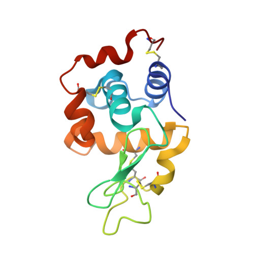
Silver metallation of hen egg white lysozyme: X-ray crystal structure and NMR studies.

Panzner, M.J.Bilinovich, S.M.Youngs, W.J.Leeper, T.C.

(2011) Chem Commun (Camb) 47: 12479-12481

  • DOI: https://doi.org/10.1039/c1cc15908a
  • Primary Citation Related Structures: 
    3RU5

  • PubMed Abstract: 

    The X-ray crystal structure, NMR binding studies, and enzyme activity of silver(I) metallated hen egg white lysozyme are presented. Primary bonding of silver is observed through His15 with secondary bonding interactions coming from nearby Arg14 and Asp87. A covalently bound nitrate completes a four coordinate binding pocket.


  • Organizational Affiliation
    • Center for Silver Therapeutics Research, Department of Chemistry, The University of Akron, Akron, OH 44325-3601, USA.

Macromolecule Content 

  • Total Structure Weight: 15.05 kDa 
  • Atom Count: 1,192 
  • Modeled Residue Count: 129 
  • Deposited Residue Count: 129 
  • Unique protein chains: 1

Macromolecules

Find similar proteins by:|  3D Structure
Entity ID: 1
MoleculeChains  Sequence LengthOrganismDetailsImage
Lysozyme C129Gallus gallusMutation(s): 0 
EC: 3.2.1.17
UniProt
Find proteins for P00698 (Gallus gallus)
Explore P00698 
Go to UniProtKB:  P00698
Entity Groups
Sequence Clusters30% Identity50% Identity70% Identity90% Identity95% Identity100% Identity
UniProt GroupP00698
Sequence Annotations
Expand
Reference Sequence

Small Molecules

Ligands 4 Unique
IDChains Name / Formula / InChI Key2D Diagram3D Interactions
AG

Query on AG


Download:Ideal Coordinates CCD File
B [auth A]SILVER ION
Ag
FOIXSVOLVBLSDH-UHFFFAOYSA-N
EDO

Query on EDO


Download:Ideal Coordinates CCD File
J [auth A],
M [auth A]
1,2-ETHANEDIOL
C2 H6 O2
LYCAIKOWRPUZTN-UHFFFAOYSA-N
NO3

Query on NO3


Download:Ideal Coordinates CCD File
C [auth A]
D [auth A]
K [auth A]
L [auth A]
N [auth A]
C [auth A],
D [auth A],
K [auth A],
L [auth A],
N [auth A],
O [auth A]
NITRATE ION
N O3
NHNBFGGVMKEFGY-UHFFFAOYSA-N
NA

Query on NA


Download:Ideal Coordinates CCD File
E [auth A],
F [auth A],
G [auth A],
H [auth A],
I [auth A]
SODIUM ION
Na
FKNQFGJONOIPTF-UHFFFAOYSA-N

Experimental Data & Validation

Experimental Data

  • Method: X-RAY DIFFRACTION
  • Resolution: 1.35 Å
  • R-Value Free:  0.215 (Depositor), 0.216 (DCC) 
  • R-Value Work:  0.192 (Depositor), 0.194 (DCC) 
  • R-Value Observed: 0.193 (Depositor) 
Space Group: P 43 21 2
Unit Cell:
Length ( Å )Angle ( ˚ )
a = 77.798α = 90
b = 77.798β = 90
c = 37.629γ = 90
Software Package:
Software NamePurpose
APEXdata collection
PHASERphasing
REFMACrefinement
SAINTdata reduction
APEXdata reduction

Structure Validation

View Full Validation Report


Entry History 

Deposition Data

Revision History  (Full details and data files)

  • Version 1.0: 2011-11-23
    Type: Initial release
  • Version 1.1: 2018-04-04
    Changes: Data collection
  • Version 1.2: 2023-09-13
    Changes: Data collection, Database references, Derived calculations, Refinement description
  • Version 1.3: 2024-11-27
    Changes: Structure summary