3RKX | pdb_00003rkx

Structural characterisation of staphylococcus aureus biotin protein ligase


Experimental Data Snapshot

  • Method: X-RAY DIFFRACTION
  • Resolution: 2.10 Å
  • R-Value Free: 
    0.266 (Depositor), 0.262 (DCC) 
  • R-Value Work: 
    0.193 (Depositor), 0.188 (DCC) 
  • R-Value Observed: 
    0.197 (Depositor) 

Starting Model: experimental
View more details

wwPDB Validation 3D Report Full Report

Validation slider image for 3RKX

This is version 1.1 of the entry. See complete history

Literature

Structural characterisation of staphylococcus aureus biotin protein ligase

Pendini, N.R.Yap, M.Y.Polyak, S.W.Cowieson, N.Traore, D.A.K.Booker, G.W.Wallace, J.C.Abell, A.Wilce, J.A.Wilce, M.C.J.

To be published.

Macromolecule Content 

  • Total Structure Weight: 37.19 kDa 
  • Atom Count: 2,696 
  • Modeled Residue Count: 308 
  • Deposited Residue Count: 323 
  • Unique protein chains: 1

Macromolecules

Find similar proteins by:|  3D Structure
Entity ID: 1
MoleculeChains  Sequence LengthOrganismDetailsImage
Biotin-[acetyl-CoA-carboxylase] ligase323Staphylococcus aureus subsp. aureus ECT-R 2Mutation(s): 0 
Gene Names: ECTR2_1310
EC: 6.3.4.15
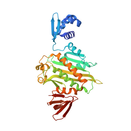
Experimental Data & Validation

Experimental Data

  • Method: X-RAY DIFFRACTION
  • Resolution: 2.10 Å
  • R-Value Free:  0.266 (Depositor), 0.262 (DCC) 
  • R-Value Work:  0.193 (Depositor), 0.188 (DCC) 
  • R-Value Observed: 0.197 (Depositor) 
Space Group: P 1 21 1
Unit Cell:
Length ( Å )Angle ( ˚ )
a = 50.132α = 90
b = 51.401β = 108
c = 67.584γ = 90
Software Package:
Software NamePurpose
Blu-Icedata collection
PHASERphasing
PHENIXrefinement
MOSFLMdata reduction
SCALAdata scaling

Structure Validation

View Full Validation Report


Entry History 

Deposition Data

Revision History  (Full details and data files)

  • Version 1.0: 2012-04-18
    Type: Initial release
  • Version 1.1: 2023-11-01
    Changes: Data collection, Database references, Refinement description