3RKF | pdb_00003rkf

Crystal structure of guanine riboswitch C61U/G37A double mutant bound to thio-guanine


Experimental Data Snapshot

  • Method: X-RAY DIFFRACTION
  • Resolution: 2.50 Å
  • R-Value Free: 
    0.226 (Depositor), 0.214 (DCC) 
  • R-Value Work: 
    0.207 (Depositor), 0.204 (DCC) 
  • R-Value Observed: 
    0.208 (Depositor) 

Starting Model: experimental
View more details

wwPDB Validation 3D Report Full Report

Validation slider image for 3RKF

Ligand Structure Quality Assessment 


This is version 1.3 of the entry. See complete history

Literature

Influence of ground-state structure and Mg2+ binding on folding kinetics of the guanine-sensing riboswitch aptamer domain.

Buck, J.Wacker, A.Warkentin, E.Wohnert, J.Wirmer-Bartoschek, J.Schwalbe, H.

(2011) Nucleic Acids Res 39: 9768-9778

  • DOI: https://doi.org/10.1093/nar/gkr664
  • Primary Citation Related Structures: 
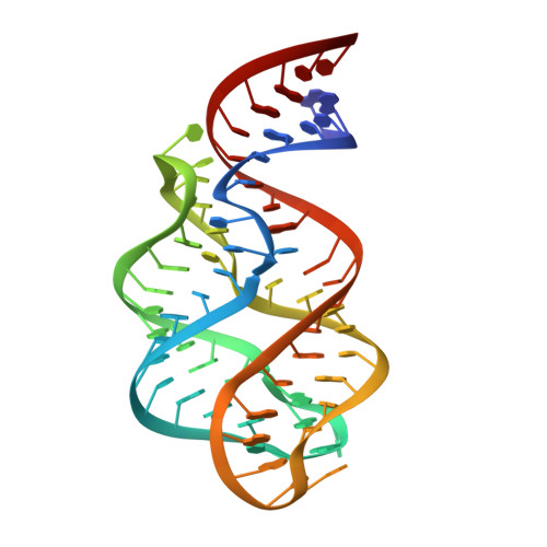
    3RKF

  • PubMed Abstract: 

    Riboswitch RNAs fold into complex tertiary structures upon binding to their cognate ligand. Ligand recognition is accomplished by key residues in the binding pocket. In addition, it often crucially depends on the stability of peripheral structural elements. The ligand-bound complex of the guanine-sensing riboswitch from Bacillus subtilis, for example, is stabilized by extensive interactions between apical loop regions of the aptamer domain. Previously, we have shown that destabilization of this tertiary loop-loop interaction abrogates ligand binding of the G37A/C61U-mutant aptamer domain (Gsw(loop)) in the absence of Mg(2+). However, if Mg(2+) is available, ligand-binding capability is restored by a population shift of the ground-state RNA ensemble toward RNA conformations with pre-formed loop-loop interactions. Here, we characterize the striking influence of long-range tertiary structure on RNA folding kinetics and on ligand-bound complex structure, both by X-ray crystallography and time-resolved NMR. The X-ray structure of the ligand-bound complex reveals that the global architecture is almost identical to the wild-type aptamer domain. The population of ligand-binding competent conformations in the ground-state ensemble of Gsw(loop) is tunable through variation of the Mg(2+) concentration. We quantitatively describe the influence of distinct Mg(2+) concentrations on ligand-induced folding trajectories both by equilibrium and time-resolved NMR spectroscopy at single-residue resolution.


  • Organizational Affiliation
    • Institute for Organic Chemistry and Chemical Biology, Center for Biomolecular Magnetic Resonance, Johann Wolfgang Goethe-University, Max-von-Laue-Strasse 7 & 9, 60438 Frankfurt am Main, Germany.

Macromolecule Content 

  • Total Structure Weight: 90.95 kDa 
  • Atom Count: 5,946 
  • Modeled Residue Count: 268 
  • Deposited Residue Count: 268 
  • Unique nucleic acid chains: 1

Macromolecules

Find similar nucleic acids by:  (by identity cutoff) 
Entity ID: 1
MoleculeChains LengthOrganismImage
Guanine riboswitch
A, B, C, D
67N/A
Sequence Annotations
Expand
Reference Sequence

Small Molecules

Ligands 2 Unique
IDChains Name / Formula / InChI Key2D Diagram3D Interactions
DX4

Query on DX4


Download:Ideal Coordinates CCD File
AA [auth D],
E [auth A],
O [auth B],
U [auth C]
2-amino-1,9-dihydro-6H-purine-6-thione
C5 H5 N5 S
WYWHKKSPHMUBEB-UHFFFAOYSA-N
NCO

Query on NCO


Download:Ideal Coordinates CCD File
BA [auth D]
CA [auth D]
DA [auth D]
EA [auth D]
F [auth A]
BA [auth D],
CA [auth D],
DA [auth D],
EA [auth D],
F [auth A],
FA [auth D],
G [auth A],
GA [auth D],
H [auth A],
HA [auth D],
I [auth A],
J [auth A],
K [auth A],
L [auth A],
M [auth A],
N [auth A],
P [auth B],
Q [auth B],
R [auth B],
S [auth B],
T [auth B],
V [auth C],
W [auth C],
X [auth C],
Y [auth C],
Z [auth C]
COBALT HEXAMMINE(III)
Co H18 N6
DYLMFCCYOUSRTK-UHFFFAOYSA-N

Experimental Data & Validation

Experimental Data

  • Method: X-RAY DIFFRACTION
  • Resolution: 2.50 Å
  • R-Value Free:  0.226 (Depositor), 0.214 (DCC) 
  • R-Value Work:  0.207 (Depositor), 0.204 (DCC) 
  • R-Value Observed: 0.208 (Depositor) 
Space Group: P 32
Unit Cell:
Length ( Å )Angle ( ˚ )
a = 52.3α = 90
b = 52.3β = 90
c = 263.4γ = 120
Software Package:
Software NamePurpose
PHASERphasing
PHENIXrefinement
XDSdata reduction
XSCALEdata scaling

Structure Validation

View Full Validation Report


Ligand Structure Quality Assessment 


Entry History 

Deposition Data

Revision History  (Full details and data files)

  • Version 1.0: 2011-08-17
    Type: Initial release
  • Version 1.1: 2011-10-26
    Changes: Database references
  • Version 1.2: 2011-12-28
    Changes: Database references
  • Version 1.3: 2023-09-13
    Changes: Data collection, Database references, Derived calculations, Refinement description