


Crystal structure of the DNA binding domain of CovR from Streptococcus pyogenes
Horstmann, N., Kumaraswami, M., Musser, J.M., Brennan, R.G., Shelburne, S.A.To be published.
Experimental Data Snapshot
wwPDB Validation 3D Report Full Report

Entity ID: 1 | |||||
|---|---|---|---|---|---|
| Molecule | Chains | Sequence Length | Organism | Details | Image |
| CovR | 96 | Streptococcus pyogenes | Mutation(s): 0 Gene Names: covR, csrR | 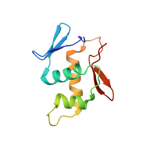 | |
UniProt | |||||
Entity Groups | |||||
| Sequence Clusters | 30% Identity50% Identity70% Identity90% Identity95% Identity100% Identity | ||||
| UniProt Group | O87527 | ||||
Sequence AnnotationsExpand | |||||
Reference Sequence | |||||
| Length ( Å ) | Angle ( ˚ ) |
|---|---|
| a = 30.467 | α = 90 |
| b = 36.686 | β = 94.6 |
| c = 38.937 | γ = 90 |
| Software Name | Purpose |
|---|---|
| ADSC | data collection |
| SOLVE | phasing |
| CNS | refinement |
| MOSFLM | data reduction |
| SCALA | data scaling |


