


Crystal structure and kinetic studies of carbohydrate oxidase from Microdochium nivale
Duskova, J., Skalova, T., Kolenko, P., Stepankova, A., Hasek, J., Koval, T., Ostergaard, L.H., Fuglsang, C.C., Dohnalek, J.To be published.
Experimental Data Snapshot
Starting Model: experimental
View more details
Entity ID: 1 | |||||
|---|---|---|---|---|---|
| Molecule | Chains | Sequence Length | Organism | Details | Image |
| Carbohydrate oxidase | 473 | Microdochium nivale | Mutation(s): 0 Gene Names: MnCO EC: 1.1.3.5 (PDB Primary Data), 1.1.3 (UniProt) | 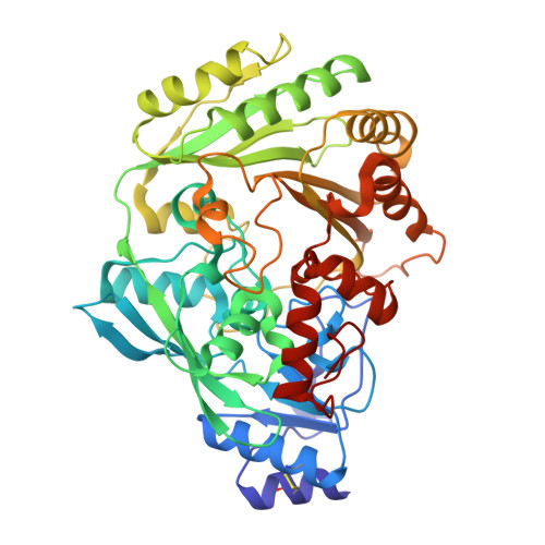 | |
UniProt | |||||
Entity Groups | |||||
| Sequence Clusters | 30% Identity50% Identity70% Identity90% Identity95% Identity100% Identity | ||||
| UniProt Group | I1SB12 | ||||
Glycosylation | |||||
| Glycosylation Sites: 1 | |||||
Sequence AnnotationsExpand | |||||
Reference Sequence | |||||
| Ligands 7 Unique | |||||
|---|---|---|---|---|---|
| ID | Chains | Name / Formula / InChI Key | 2D Diagram | 3D Interactions | |
| FAD Download:Ideal Coordinates CCD File | B [auth A] | FLAVIN-ADENINE DINUCLEOTIDE C27 H33 N9 O15 P2 VWWQXMAJTJZDQX-UYBVJOGSSA-N |  | ||
| ABL Download:Ideal Coordinates CCD File | G [auth A], H [auth A] | (2R,3R,4R,5R)-4,5-dihydroxy-2-(hydroxymethyl)-6-oxopiperidin-3-yl beta-D-glucopyranoside C12 H21 N O10 WXSNJJDPPISYEF-ZNLUKOTNSA-N |  | ||
| NAG Download:Ideal Coordinates CCD File | C [auth A] | 2-acetamido-2-deoxy-beta-D-glucopyranose C8 H15 N O6 OVRNDRQMDRJTHS-FMDGEEDCSA-N |  | ||
| TRS Download:Ideal Coordinates CCD File | I [auth A] | 2-AMINO-2-HYDROXYMETHYL-PROPANE-1,3-DIOL C4 H12 N O3 LENZDBCJOHFCAS-UHFFFAOYSA-O |  | ||
| SO4 Download:Ideal Coordinates CCD File | J [auth A] | SULFATE ION O4 S QAOWNCQODCNURD-UHFFFAOYSA-L |  | ||
| ZN Download:Ideal Coordinates CCD File | D [auth A], E [auth A], F [auth A] | ZINC ION Zn PTFCDOFLOPIGGS-UHFFFAOYSA-N |  | ||
| CL Download:Ideal Coordinates CCD File | K [auth A], L [auth A], M [auth A], N [auth A] | CHLORIDE ION Cl VEXZGXHMUGYJMC-UHFFFAOYSA-M |  | ||
| Length ( Å ) | Angle ( ˚ ) |
|---|---|
| a = 132.04 | α = 90 |
| b = 56.92 | β = 95.55 |
| c = 86.9 | γ = 90 |
| Software Name | Purpose |
|---|---|
| CrysalisPro | data collection |
| MOLREP | phasing |
| REFMAC | refinement |
| CrysalisPro | data reduction |
| Jana2006 | data scaling |


