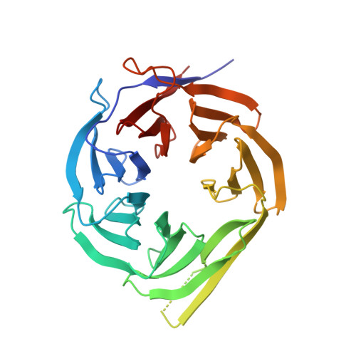
Structure of the RACK1 dimer from Saccharomyces cerevisiae
Yatime, L., Hein, K.L., Nilsson, J., Nissen, P.(2011) J Mol Biology 411: 486-498
- PubMed: 21704636
- DOI: https://doi.org/10.1016/j.jmb.2011.06.017
- Primary Citation of Related Structures:
3RFG, 3RFH - PubMed Abstract:
Receptor for activated C-kinase 1 (RACK1) serves as a scaffolding protein in numerous signaling pathways involving kinases and membrane-bound receptors from different cellular compartments. It exists simultaneously as a cytosolic free form and as a ribosome-bound protein. As part of the 40S ribosomal subunit, it triggers translational regulation by establishing a direct link between protein kinase C and the protein synthesis machinery. It has been suggested that RACK1 could recruit other signaling molecules onto the ribosome, providing a signal-specific modulation of the translational process. RACK1 is able to dimerize both in vitro and in vivo. This homodimer formation has been observed in several processes including the regulation of the N-methyl-d-aspartate receptor by the Fyn kinase in the brain and the oxygen-independent degradation of hypoxia-inducible factor 1. The functional relevance of this dimerization is, however, still unclear and the question of a possible dimerization of the ribosome-bound protein is still pending. Here, we report the first structure of a RACK1 homodimer, as determined from two independent crystal forms of the Saccharomyces cerevisiae RACK1 protein (also known as Asc1p) at 2.9 and 3.9 Å resolution. The structure reveals an atypical mode of dimerization where monomers intertwine on blade 4, thus exposing a novel surface of the protein to potential interacting partners. We discuss the significance of the dimer structure for RACK1 function.
- Department of Molecular Biology, Aarhus University, Gustav Wieds Vej 10C, DK-8000 Aarhus C, Denmark. lay@mb.au.dk
Organizational Affiliation:


