3RE0 | pdb_00003re0

Crystal structure of human apo Cu,Zn superoxide dismutase (SOD1) complexed with cisplatin


Experimental Data Snapshot

  • Method: X-RAY DIFFRACTION
  • Resolution: 2.28 Å
  • R-Value Free: 
    0.290 (Depositor), 0.293 (DCC) 
  • R-Value Work: 
    0.209 (Depositor), 0.220 (DCC) 
  • R-Value Observed: 
    0.216 (Depositor) 

Starting Model: experimental
View more details

wwPDB Validation 3D Report Full Report

Validation slider image for 3RE0

This is version 1.3 of the entry. See complete history

Literature

Interaction of cisplatin with human superoxide dismutase.

Banci, L.Bertini, I.Blazevits, O.Calderone, V.Cantini, F.Mao, J.Trapananti, A.Vieru, M.Amori, I.Cozzolino, M.Carri, M.T.

(2012) J Am Chem Soc 134: 7009-7014

  • DOI: https://doi.org/10.1021/ja211591n
  • Primary Citation Related Structures: 
    3RE0

  • PubMed Abstract: 

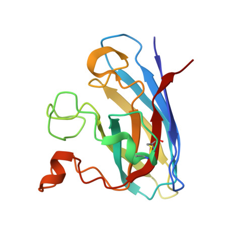
    cis-Diamminedichloroplatinum(II) (cisplatin) is able to interact with human superoxide dismutase (hSOD1) in the disulfide oxidized apo form with a dissociation constant of 37 ± 3 μM through binding cysteine 111 (Cys111) located at the edge of the subunit interface. It also binds to Cu(2)-Zn(2) and Zn(2)-Zn(2) forms of hSOD1. Cisplatin inhibits aggregation of demetalated oxidized hSOD1, and it is further able to dissolve and monomerize oxidized hSOD1 oligomers in vitro and in cell, thus indicating its potential as a leading compound for amyotrophic lateral sclerosis.


  • Organizational Affiliation
    • Magnetic Resonance Center CERM, University of Florence, Via Luigi Sacconi 6, 50019 Sesto Fiorentino, Florence, Italy. banci@cerm.unifi.it

Macromolecule Content 

  • Total Structure Weight: 64.21 kDa 
  • Atom Count: 4,143 
  • Modeled Residue Count: 555 
  • Deposited Residue Count: 612 
  • Unique protein chains: 1

Macromolecules

Find similar proteins by:|  3D Structure
Entity ID: 1
MoleculeChains  Sequence LengthOrganismDetailsImage
Superoxide dismutase [Cu-Zn]
A, B, C, D
153Homo sapiensMutation(s): 0 
Gene Names: SOD1
EC: 1.15.1.1 (PDB Primary Data), 1.8 (UniProt)
UniProt & NIH Common Fund Data Resources
Find proteins for P00441 (Homo sapiens)
Explore P00441 
Go to UniProtKB:  P00441
PHAROS:  P00441
GTEx:  ENSG00000142168 
Entity Groups
Sequence Clusters30% Identity50% Identity70% Identity90% Identity95% Identity100% Identity
UniProt GroupP00441
Sequence Annotations
Expand
Reference Sequence

Experimental Data & Validation

Experimental Data

  • Method: X-RAY DIFFRACTION
  • Resolution: 2.28 Å
  • R-Value Free:  0.290 (Depositor), 0.293 (DCC) 
  • R-Value Work:  0.209 (Depositor), 0.220 (DCC) 
  • R-Value Observed: 0.216 (Depositor) 
Space Group: C 1 2 1
Unit Cell:
Length ( Å )Angle ( ˚ )
a = 157.412α = 90
b = 34.805β = 112.28
c = 115.225γ = 90
Software Package:
Software NamePurpose
ADSCdata collection
MOLREPphasing
REFMACrefinement
MOSFLMdata reduction
SCALAdata scaling

Structure Validation

View Full Validation Report


Entry History 

Deposition Data

Revision History  (Full details and data files)

  • Version 1.0: 2012-04-25
    Type: Initial release
  • Version 1.1: 2013-07-03
    Changes: Database references
  • Version 1.2: 2023-11-01
    Changes: Data collection, Database references, Derived calculations, Refinement description
  • Version 1.3: 2024-10-30
    Changes: Structure summary