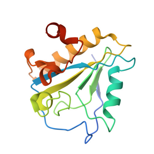
Role of net charge on catalytic domain and influence of cell wall binding domain on bactericidal activity, specificity, and host range of phage lysins.
Low, L.Y., Yang, C., Perego, M., Osterman, A., Liddington, R.(2011) J Biological Chem 286: 34391-34403
- PubMed: 21816821
- DOI: https://doi.org/10.1074/jbc.M111.244160
- Primary Citation of Related Structures:
3HMB, 3HMC, 3RDR - PubMed Abstract:
The recombinant lysins of lytic phages, when applied externally to Gram-positive bacteria, can be efficient bactericidal agents, typically retaining high specificity. Their development as novel antibacterial agents offers many potential advantages over conventional antibiotics. Protein engineering could exploit this potential further by generating novel lysins fit for distinct target populations and environments. However, access to the peptidoglycan layer is controlled by a variety of secondary cell wall polymers, chemical modifications, and (in some cases) S-layers and capsules. Classical lysins require a cell wall-binding domain (CBD) that targets the catalytic domain to the peptidoglycan layer via binding to a secondary cell wall polymer component. The cell walls of Gram-positive bacteria generally have a negative charge, and we noticed a correlation between (positive) charge on the catalytic domain and bacteriolytic activity in the absence of the CBD (nonclassical behavior). We investigated a physical basis for this correlation by comparing the structures and activities of pairs of lysins where the lytic activity of one of each pair was CBD-independent. We found that by engineering a reversal of sign of the net charge of the catalytic domain, we could either eliminate or create CBD dependence. We also provide evidence that the S-layer of Bacillus anthracis acts as a molecular sieve that is chiefly size-dependent, favoring catalytic domains over full-length lysins. Our work suggests a number of facile approaches for fine-tuning lysin activity, either to enhance or reduce specificity/host range and/or bactericidal potential, as required.
- Infectious and Inflammatory Disease Center, Sanford-Burnham Medical Research Institute, La Jolla, California 92037, USA.
Organizational Affiliation:


