3RDK | pdb_00003rdk

Protein crystal structure of xylanase A1 of Paenibacillus sp. JDR-2


Experimental Data Snapshot

  • Method: X-RAY DIFFRACTION
  • Resolution: 1.49 Å
  • R-Value Free: 
    0.184 (Depositor), 0.185 (DCC) 
  • R-Value Work: 
    0.145 (Depositor), 0.145 (DCC) 
  • R-Value Observed: 
    0.147 (Depositor) 

wwPDB Validation 3D Report Full Report

Validation slider image for 3RDK

This is version 2.1 of the entry. See complete history

Literature

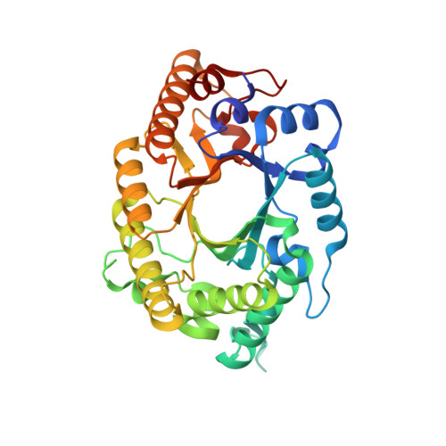
Novel structural features of xylanase A1 from Paenibacillus sp. JDR-2.

St John, F.J.Preston, J.F.Pozharski, E.

(2012) J Struct Biol 180: 303-311

  • DOI: https://doi.org/10.1016/j.jsb.2012.09.007
  • Primary Citation Related Structures: 
    3RDK, 3RO8, 4E4P

  • PubMed Abstract: 

    The Gram-positive bacterium Paenibacillus sp. JDR-2 (PbJDR2) has been shown to have novel properties in the utilization of the abundant but chemically complex hemicellulosic sugar glucuronoxylan. Xylanase A1 of PbJDR2 (PbXynA1) has been implicated in an efficient process in which extracellular depolymerization of this polysaccharide is coupled to assimilation and intracellular metabolism. PbXynA1is a 154kDa cell wall anchored multimodular glycosyl hydrolase family 10 (GH10) xylanase. In this work, the 38kDa catalytic module of PbXynA1 has been structurally characterized revealing several new features not previously observed in structures of GH10 xylanases. These features are thought to facilitate hydrolysis of highly substituted, chemically complex xylans that may be the form found in close proximity to the cell wall of PbJDR2, an organism shown to have a preference for growth on polymeric glucuronoxylan.


  • Organizational Affiliation
    • Department of Pharmaceutical Sciences, University of Maryland School of Pharmacy, 20 Penn Street, Baltimore, MD 21201, USA. fjstjohn@gmail.com

Macromolecule Content 

  • Total Structure Weight: 78.97 kDa 
  • Atom Count: 6,364 
  • Modeled Residue Count: 667 
  • Deposited Residue Count: 682 
  • Unique protein chains: 1

Macromolecules

Find similar proteins by:|  3D Structure
Entity ID: 1
MoleculeChains  Sequence LengthOrganismDetailsImage
Endo-1,4-beta-xylanase
A, B
341Paenibacillus sp. JDR-2Mutation(s): 0 
Gene Names: Pjdr2_0221
EC: 3.2.1.8
UniProt
Find proteins for C6CRV0 (Paenibacillus sp. (strain JDR-2))
Explore C6CRV0 
Go to UniProtKB:  C6CRV0
Entity Groups
Sequence Clusters30% Identity50% Identity70% Identity90% Identity95% Identity100% Identity
UniProt GroupC6CRV0
Sequence Annotations
Expand
Reference Sequence

Oligosaccharides

Help  
Entity ID: 2
MoleculeChains Length2D Diagram GlycosylationD Interactions
4-O-methyl-alpha-D-glucopyranuronic acid-(1-2)-beta-D-xylopyranose-(1-4)-beta-D-xylopyranose-(1-4)-alpha-D-xylopyranose
C, D
4N/A
Glycosylation Resources
GlyTouCan: G82480QZ
GlyCosmos: G82480QZ

Small Molecules

Ligands 3 Unique
IDChains Name / Formula / InChI Key2D Diagram3D Interactions
GOL

Query on GOL


Download:Ideal Coordinates CCD File
E [auth A]
F [auth A]
H [auth A]
K [auth A]
L [auth A]
E [auth A],
F [auth A],
H [auth A],
K [auth A],
L [auth A],
N [auth B],
O [auth B],
P [auth B],
Q [auth B]
GLYCEROL
C3 H8 O3
PEDCQBHIVMGVHV-UHFFFAOYSA-N
CL

Query on CL


Download:Ideal Coordinates CCD File
G [auth A],
I [auth A],
J [auth A]
CHLORIDE ION
Cl
VEXZGXHMUGYJMC-UHFFFAOYSA-M
MG

Query on MG


Download:Ideal Coordinates CCD File
M [auth A]MAGNESIUM ION
Mg
JLVVSXFLKOJNIY-UHFFFAOYSA-N

Experimental Data & Validation

Experimental Data

  • Method: X-RAY DIFFRACTION
  • Resolution: 1.49 Å
  • R-Value Free:  0.184 (Depositor), 0.185 (DCC) 
  • R-Value Work:  0.145 (Depositor), 0.145 (DCC) 
  • R-Value Observed: 0.147 (Depositor) 
Space Group: P 43 21 2
Unit Cell:
Length ( Å )Angle ( ˚ )
a = 165.113α = 90
b = 165.113β = 90
c = 66.355γ = 90
Software Package:
Software NamePurpose
HKL-2000data collection
PHASERphasing
REFMACrefinement
HKL-2000data scaling

Structure Validation

View Full Validation Report


Entry History 

Deposition Data

Revision History  (Full details and data files)

  • Version 1.0: 2012-04-04
    Type: Initial release
  • Version 1.1: 2012-10-10
    Changes: Database references
  • Version 1.2: 2012-11-14
    Changes: Database references
  • Version 1.3: 2017-10-25
    Changes: Author supporting evidence
  • Version 2.0: 2020-07-29
    Type: Remediation
    Reason: Carbohydrate remediation
    Changes: Advisory, Atomic model, Data collection, Database references, Derived calculations, Structure summary
  • Version 2.1: 2024-02-21
    Changes: Data collection, Database references, Structure summary