3R3M | pdb_00003r3m

Crystal structure of the FAF1 UBX domain


Experimental Data Snapshot

  • Method: X-RAY DIFFRACTION
  • Resolution: 3.00 Å
  • R-Value Free: 
    0.258 (Depositor), 0.255 (DCC) 
  • R-Value Work: 
    0.174 (Depositor), 0.175 (DCC) 
  • R-Value Observed: 
    0.178 (Depositor) 

Starting Model: experimental
View more details

wwPDB Validation 3D Report Full Report

Validation slider image for 3R3M

This is version 1.3 of the entry. See complete history

Literature

Hierarchical Binding of Cofactors to the AAA ATPase p97.

Hanzelmann, P.Buchberger, A.Schindelin, H.

(2011) Structure 19: 833-843

  • DOI: https://doi.org/10.1016/j.str.2011.03.018
  • Primary Citation Related Structures: 
    3QQ7, 3QQ8, 3R3M

  • PubMed Abstract: 

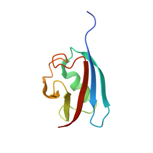
    The hexameric AAA ATPase p97 is involved in several human proteinopathies and mediates ubiquitin-dependent protein degradation among other essential cellular processes. Via its N-terminal domain (N domain), p97 interacts with multiple regulatory cofactors including the UFD1/NPL4 heterodimer and members of the "ubiquitin regulatory X" (UBX) domain protein family; however, the principles governing cofactor selectivity remain to be deciphered. Our crystal structure of the FAS-associated factor 1 (FAF1)UBX domain in complex with the p97N domain reveals that the signature Phe-Pro-Arg motif known to be crucial for interactions of UBX domains with p97 adopts a cis-proline configuration, in contrast to a cis-trans mixture we derive for the isolated FAF1UBX domain. Biochemical studies confirm that binding critically depends on a proline at this position. Furthermore, we observe that the UBX proteins FAF1 and UBXD7 only bind to p97-UFD1/NPL4, but not free p97, thus demonstrating for the first time a hierarchy in p97-cofactor interactions.


  • Organizational Affiliation
    • Rudolf Virchow Center for Experimental Biomedicine, University of Würzburg, Josef-Schneider-Str. 2, 97080 Würzburg, Germany. petra.haenzelmann@virchow.uni-wuerzburg.de

Macromolecule Content 

  • Total Structure Weight: 40.07 kDa 
  • Atom Count: 2,763 
  • Modeled Residue Count: 330 
  • Deposited Residue Count: 340 
  • Unique protein chains: 1

Macromolecules

Find similar proteins by:|  3D Structure
Entity ID: 1
MoleculeChains  Sequence LengthOrganismDetailsImage
FAS-associated factor 1A [auth B],
B [auth A],
C,
D
85Homo sapiensMutation(s): 0 
Gene Names: FAF1UBXD12UBXN3ACGI-03
UniProt & NIH Common Fund Data Resources
Find proteins for Q9UNN5 (Homo sapiens)
Explore Q9UNN5 
Go to UniProtKB:  Q9UNN5
PHAROS:  Q9UNN5
GTEx:  ENSG00000185104 
Entity Groups
Sequence Clusters30% Identity50% Identity70% Identity90% Identity95% Identity100% Identity
UniProt GroupQ9UNN5
Sequence Annotations
Expand
Reference Sequence

Experimental Data & Validation

Experimental Data

  • Method: X-RAY DIFFRACTION
  • Resolution: 3.00 Å
  • R-Value Free:  0.258 (Depositor), 0.255 (DCC) 
  • R-Value Work:  0.174 (Depositor), 0.175 (DCC) 
  • R-Value Observed: 0.178 (Depositor) 
Space Group: P 61
Unit Cell:
Length ( Å )Angle ( ˚ )
a = 92.755α = 90
b = 92.755β = 90
c = 108.5γ = 120
Software Package:
Software NamePurpose
PHENIXrefinement
PDB_EXTRACTdata extraction
MxCuBEdata collection
MOSFLMdata reduction
SCALAdata scaling
PHASERphasing
REFMACrefinement

Structure Validation

View Full Validation Report


Entry History 

Deposition Data

Revision History  (Full details and data files)

  • Version 1.0: 2011-06-22
    Type: Initial release
  • Version 1.1: 2011-07-13
    Changes: Version format compliance
  • Version 1.2: 2019-07-17
    Changes: Data collection, Refinement description
  • Version 1.3: 2023-09-13
    Changes: Data collection, Database references, Derived calculations, Refinement description