


Crystal structure of a carbonic anhydrase from a crude oil degrading psychrophilic library
Petit, P., Xu, X., Cui, H., Brown, G., Dong, A., Savchenko, A., Yakunin, A.F.To be published.
Experimental Data Snapshot
Starting Model: experimental
View more details
wwPDB Validation 3D Report Full Report

Entity ID: 1 | |||||
|---|---|---|---|---|---|
| Molecule | Chains | Sequence Length | Organism | Details | Image |
| carbonic anhydrase | 189 | unidentified | Mutation(s): 0 | 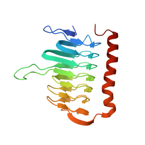 | |
| Length ( Å ) | Angle ( ˚ ) |
|---|---|
| a = 54.071 | α = 90 |
| b = 89.943 | β = 96.37 |
| c = 54.72 | γ = 90 |
| Software Name | Purpose |
|---|---|
| CrystalClear | data collection |
| MOLREP | phasing |
| PHENIX | refinement |
| XDS | data reduction |
| SCALA | data scaling |


