3QRX | pdb_00003qrx

Chlamydomonas reinhardtii centrin bound to melittin


Experimental Data Snapshot

  • Method: X-RAY DIFFRACTION
  • Resolution: 2.20 Å
  • R-Value Free: 
    0.341 (Depositor), 0.333 (DCC) 
  • R-Value Work: 
    0.291 (Depositor), 0.285 (DCC) 
  • R-Value Observed: 
    0.294 (Depositor) 

Starting Model: experimental
View more details

wwPDB Validation 3D Report Full Report

Validation slider image for 3QRX

This is version 1.2 of the entry. See complete history

Literature

The structure, molecular dynamics, and energetics of centrin-melittin complex.

Sosa Ldel, V.Alfaro, E.Santiago, J.Narvaez, D.Rosado, M.C.Rodriguez, A.Gomez, A.M.Schreiter, E.R.Pastrana-Rios, B.

(2011) Proteins 79: 3132-3143

  • DOI: https://doi.org/10.1002/prot.23142
  • Primary Citation Related Structures: 
    3QRX

  • PubMed Abstract: 

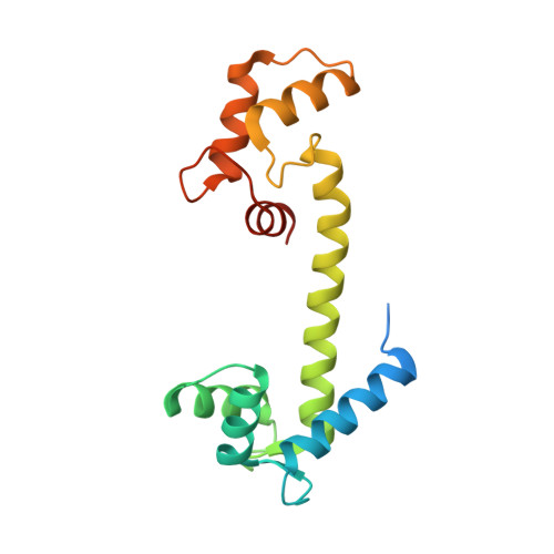
    Centrin is a calcium binding protein (CaBP) belonging to the EF-hand superfamily. As with other proteins within this family, centrin is a calcium sensor with multiple biological target proteins. We chose to study Chlamydomonas reinhardtii centrin (Crcen) and its interaction with melittin (MLT) as a model for CaBP complexes due to its amphipathic properties. Our goal was to determine the molecular interactions that lead to centrin-MLT complex formation, their relative stability, and the conformational changes associated with the interaction, when compared to the single components. For this, we determined the thermodynamic parameters that define Crcen-MLT complex formation. Two-dimensional infrared (2D IR) correlation spectroscopy were used to study the amide I', I'*, and side chain bands for (13)C-Crcen, MLT, and the (13)C-Crcen-MLT complex. This approach resulted in the determination of MLT's increased helicity, while centrin was stabilized within the complex. Herein we provide the first complete molecular description of centrin-MLT complex formation and the dissociation process. Also, discussed is the first structure of a CaBP-MLT complex by X-ray crystallography, which shows that MLT has a different binding orientation than previously characterized centrin-bound peptides. Finally, all of the experimental results presented herein are consistent with centrin maintaining an extended conformation while interacting with MLT. The molecular implications of these results are: (1) the recognition of hydrophobic contacts as requirements for initial binding, (2) minimum electrostatic interactions within the C-terminal end of the peptide, and (3) van der Waals interactions within MLTs N-terminal end are required for complex formation.


  • Organizational Affiliation
    • Department of Chemistry, University of Puerto Rico, Mayagüez, Puerto Rico 00681-9019.

Macromolecule Content 

  • Total Structure Weight: 22.5 kDa 
  • Atom Count: 1,328 
  • Modeled Residue Count: 165 
  • Deposited Residue Count: 195 
  • Unique protein chains: 2

Macromolecules

Find similar proteins by:|  3D Structure
Entity ID: 1
MoleculeChains  Sequence LengthOrganismDetailsImage
Centrin169Chlamydomonas reinhardtiiMutation(s): 0 
Gene Names: VFL2CHLREDRAFT_159554
UniProt
Find proteins for P05434 (Chlamydomonas reinhardtii)
Explore P05434 
Go to UniProtKB:  P05434
Entity Groups
Sequence Clusters30% Identity50% Identity70% Identity90% Identity95% Identity100% Identity
UniProt GroupP05434
Sequence Annotations
Expand
Reference Sequence
Find similar proteins by:|  3D Structure
Entity ID: 2
MoleculeChains  Sequence LengthOrganismDetailsImage
Melittin26Apis melliferaMutation(s): 0 
UniProt
Find proteins for P01501 (Apis mellifera)
Explore P01501 
Go to UniProtKB:  P01501
Entity Groups
Sequence Clusters30% Identity50% Identity70% Identity90% Identity95% Identity100% Identity
UniProt GroupP01501
Sequence Annotations
Expand
Reference Sequence

Experimental Data & Validation

Experimental Data

  • Method: X-RAY DIFFRACTION
  • Resolution: 2.20 Å
  • R-Value Free:  0.341 (Depositor), 0.333 (DCC) 
  • R-Value Work:  0.291 (Depositor), 0.285 (DCC) 
  • R-Value Observed: 0.294 (Depositor) 
Space Group: P 21 21 2
Unit Cell:
Length ( Å )Angle ( ˚ )
a = 52.104α = 90
b = 114.426β = 90
c = 34.842γ = 90
Software Package:
Software NamePurpose
CrystalCleardata collection
PHASERphasing
REFMACrefinement
HKL-2000data reduction
HKL-2000data scaling

Structure Validation

View Full Validation Report


Entry History 

Deposition Data

Revision History  (Full details and data files)

  • Version 1.0: 2011-10-26
    Type: Initial release
  • Version 1.1: 2013-03-20
    Changes: Derived calculations
  • Version 1.2: 2023-09-13
    Changes: Data collection, Database references, Derived calculations, Refinement description