3QPK | pdb_00003qpk

Probing oxygen channels in Melanocarpus albomyces laccase


Experimental Data Snapshot

  • Method: X-RAY DIFFRACTION
  • Resolution: 1.90 Å
  • R-Value Free: 
    0.270 (Depositor), 0.268 (DCC) 
  • R-Value Work: 
    0.214 (Depositor), 0.212 (DCC) 
  • R-Value Observed: 
    0.217 (Depositor) 

Starting Model: experimental
View more details

wwPDB Validation 3D Report Full Report

Validation slider image for 3QPK

Ligand Structure Quality Assessment 


This is version 2.2 of the entry. See complete history

Literature

Probing the Dioxygen Route in Melanocarpus albomyces Laccase with Pressurized Xenon Gas.

Kallio, J.P.Rouvinen, J.Kruus, K.Hakulinen, N.

(2011) Biochemistry 50: 4396-4398

  • DOI: https://doi.org/10.1021/bi200486b
  • Primary Citation Related Structures: 
    3QPK

  • PubMed Abstract: 

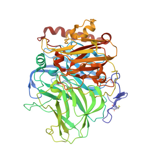
    Laccases catalyze the oxidation of phenolic substrates and the concominant reduction of dioxygen to water. We used xenon as an oxygen probe in search of routes for the entry of dioxygen into the catalytic center. Two xenon-pressurized crystal structures of recombinant Melanocarpus albomyces laccase were determined, showing three hydrophobic Xe-binding sites located in domain C. The analysis of hydrophobic cavities in other laccase structures further suggested the preference of domain C for binding of hydrophobic species such as dioxygen, thus suggesting that the hydrophobic core of domain C could function as a channel through which dioxygen can enter the trinuclear copper center.


  • Organizational Affiliation
    • Department of Chemistry, University of Eastern Finland, Joensuu, Finland.

Macromolecule Content 

  • Total Structure Weight: 129.33 kDa 
  • Atom Count: 10,254 
  • Modeled Residue Count: 1,118 
  • Deposited Residue Count: 1,118 
  • Unique protein chains: 1

Macromolecules

Find similar proteins by:|  3D Structure
Entity ID: 1
MoleculeChains  Sequence LengthOrganismDetailsImage
Laccase-1
A, B
559Melanocarpus albomycesMutation(s): 0 
Gene Names: LAC1
EC: 1.10.3.2
UniProt
Find proteins for Q70KY3 (Melanocarpus albomyces)
Explore Q70KY3 
Go to UniProtKB:  Q70KY3
Entity Groups
Sequence Clusters30% Identity50% Identity70% Identity90% Identity95% Identity100% Identity
UniProt GroupQ70KY3
Glycosylation
Glycosylation Sites: 6
Sequence Annotations
Expand
Reference Sequence

Oligosaccharides

Help  
Entity ID: 2
MoleculeChains Length2D Diagram GlycosylationD Interactions
2-acetamido-2-deoxy-beta-D-glucopyranose-(1-4)-2-acetamido-2-deoxy-beta-D-glucopyranose
C, D, E, F, G
C, D, E, F, G, H, I
2N-Glycosylation
Glycosylation Resources
GlyTouCan: G42666HT
GlyCosmos: G42666HT
GlyGen: G42666HT

Small Molecules

Ligands 5 Unique
IDChains Name / Formula / InChI Key2D Diagram3D Interactions
NAG

Query on NAG


Download:Ideal Coordinates CCD File
AA [auth B],
BA [auth B],
CA [auth B],
O [auth A],
P [auth A]
2-acetamido-2-deoxy-beta-D-glucopyranose
C8 H15 N O6
OVRNDRQMDRJTHS-FMDGEEDCSA-N
XE

Query on XE


Download:Ideal Coordinates CCD File
DA [auth B]
EA [auth B]
FA [auth B]
Q [auth A]
R [auth A]
DA [auth B],
EA [auth B],
FA [auth B],
Q [auth A],
R [auth A],
S [auth A]
XENON
Xe
FHNFHKCVQCLJFQ-UHFFFAOYSA-N
SO4

Query on SO4


Download:Ideal Coordinates CCD File
T [auth A],
U [auth A]
SULFATE ION
O4 S
QAOWNCQODCNURD-UHFFFAOYSA-L
CU

Query on CU


Download:Ideal Coordinates CCD File
J [auth A]
K [auth A]
L [auth A]
M [auth A]
V [auth B]
J [auth A],
K [auth A],
L [auth A],
M [auth A],
V [auth B],
W [auth B],
X [auth B],
Y [auth B]
COPPER (II) ION
Cu
JPVYNHNXODAKFH-UHFFFAOYSA-N
CL

Query on CL


Download:Ideal Coordinates CCD File
N [auth A],
Z [auth B]
CHLORIDE ION
Cl
VEXZGXHMUGYJMC-UHFFFAOYSA-M

Experimental Data & Validation

Experimental Data

  • Method: X-RAY DIFFRACTION
  • Resolution: 1.90 Å
  • R-Value Free:  0.270 (Depositor), 0.268 (DCC) 
  • R-Value Work:  0.214 (Depositor), 0.212 (DCC) 
  • R-Value Observed: 0.217 (Depositor) 
Space Group: C 1 2 1
Unit Cell:
Length ( Å )Angle ( ˚ )
a = 174.15α = 90
b = 60.2β = 99.79
c = 114.44γ = 90
Software Package:
Software NamePurpose
DNAdata collection
PHASERphasing
PHENIXrefinement
XDSdata reduction
XDSdata scaling

Structure Validation

View Full Validation Report


Ligand Structure Quality Assessment 


Entry History 

Deposition Data

Revision History  (Full details and data files)

  • Version 1.0: 2011-05-11
    Type: Initial release
  • Version 1.1: 2011-07-13
    Changes: Version format compliance
  • Version 2.0: 2020-07-29
    Type: Remediation
    Reason: Carbohydrate remediation
    Changes: Atomic model, Data collection, Derived calculations, Structure summary
  • Version 2.1: 2023-09-13
    Changes: Data collection, Database references, Refinement description, Structure summary
  • Version 2.2: 2024-11-06
    Changes: Structure summary