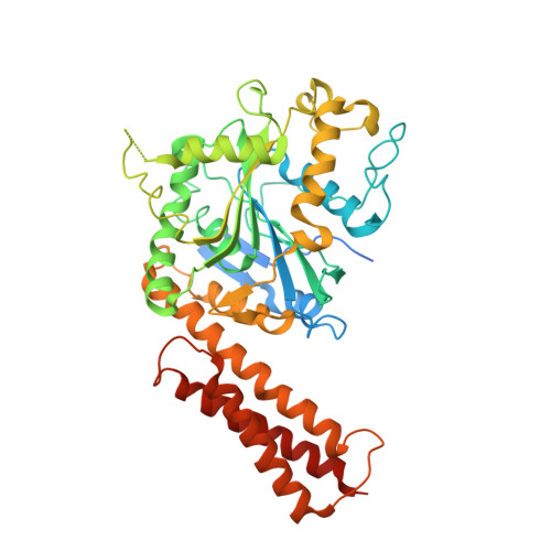
3QOF

Crystal structure of the cytosolic domain of human atlastin-1 in complex with GDP, orthorhombic form


Experimental Data Snapshot

  • Method: X-RAY DIFFRACTION
  • Resolution: 2.80 Å
  • R-Value Free: 0.267 
  • R-Value Work: 0.225 
  • R-Value Observed: 0.227 

Starting Model: experimental
View more details

wwPDB Validation   3D Report Full Report


Ligand Structure Quality Assessment 


This is version 1.2 of the entry. See complete history


Literature

Structures of the atlastin GTPase provide insight into homotypic fusion of endoplasmic reticulum membranes.

Bian, X.Klemm, R.W.Liu, T.Y.Zhang, M.Sun, S.Sui, X.Liu, X.Rapoport, T.A.Hu, J.

(2011) Proc Natl Acad Sci U S A 108: 3976-3981

  • DOI: https://doi.org/10.1073/pnas.1101643108
  • Primary Citation of Related Structures:  
    3QNU, 3QOF

  • PubMed Abstract: 

    The generation of the tubular network of the endoplasmic reticulum (ER) requires homotypic membrane fusion that is mediated by the dynamin-like, membrane-bound GTPase atlastin (ATL). Here, we have determined crystal structures of the cytosolic segment of human ATL1, which give insight into the mechanism of membrane fusion. The structures reveal a GTPase domain and athree-helix bundle, connected by a linker region. One structure corresponds to a prefusion state, in which ATL molecules in apposing membranes interact through their GTPase domains to form a dimer with the nucleotides bound at the interface. The other structure corresponds to a postfusion state generated after GTP hydrolysis and phosphate release. Compared with the prefusion structure, the three-helix bundles of the two ATL molecules undergo a major conformational change relative to the GTPase domains, which could pull the membranes together. The proposed fusion mechanism is supported by biochemical experiments and fusion assays with wild-type and mutant full-length Drosophila ATL. These experiments also show that membrane fusion is facilitated by the C-terminal cytosolic tails following the two transmembrane segments. Finally, our results show that mutations in ATL1 causing hereditary spastic paraplegia compromise homotypic ER fusion.


  • Organizational Affiliation

    Department of Genetics and Cell Biology, College of Life Sciences, and Tianjin Key Laboratory of Protein Sciences, Nankai University, Tianjin 300071, China.


Macromolecules
Find similar proteins by:  (by identity cutoff)  |  3D Structure
Entity ID: 1
MoleculeChains Sequence LengthOrganismDetailsImage
Atlastin-1
A, B, C, D
459Homo sapiensMutation(s): 0 
Gene Names: ATL1GBP3SPG3A
EC: 3.6.5
UniProt & NIH Common Fund Data Resources
Find proteins for Q8WXF7 (Homo sapiens)
Explore Q8WXF7 
Go to UniProtKB:  Q8WXF7
PHAROS:  Q8WXF7
GTEx:  ENSG00000198513 
Entity Groups  
Sequence Clusters30% Identity50% Identity70% Identity90% Identity95% Identity100% Identity
UniProt GroupQ8WXF7
Sequence Annotations
Expand
  • Reference Sequence
Small Molecules
Ligands 2 Unique
IDChains Name / Formula / InChI Key2D Diagram3D Interactions
GDP
Query on GDP

Download Ideal Coordinates CCD File 
E [auth A],
G [auth B],
I [auth C],
K [auth D]
GUANOSINE-5'-DIPHOSPHATE
C10 H15 N5 O11 P2
QGWNDRXFNXRZMB-UUOKFMHZSA-N
MG
Query on MG

Download Ideal Coordinates CCD File 
F [auth A],
H [auth B],
J [auth C],
L [auth D]
MAGNESIUM ION
Mg
JLVVSXFLKOJNIY-UHFFFAOYSA-N
Experimental Data & Validation

Experimental Data

  • Method: X-RAY DIFFRACTION
  • Resolution: 2.80 Å
  • R-Value Free: 0.267 
  • R-Value Work: 0.225 
  • R-Value Observed: 0.227 
  • Space Group: P 21 21 21
Unit Cell:
Length ( Å )Angle ( ˚ )
a = 104.742α = 90
b = 133.445β = 90
c = 176.266γ = 90
Software Package:
Software NamePurpose
HKL-2000data collection
AMoREphasing
PHENIXrefinement
HKL-2000data reduction
HKL-2000data scaling

Structure Validation

View Full Validation Report


Ligand Structure Quality Assessment 


Entry History 

Deposition Data

  • Released Date: 2011-03-02 
  • Deposition Author(s): Liu, X.

Revision History  (Full details and data files)

  • Version 1.0: 2011-03-02
    Type: Initial release
  • Version 1.1: 2011-07-13
    Changes: Version format compliance
  • Version 1.2: 2023-11-01
    Changes: Data collection, Database references, Derived calculations, Refinement description