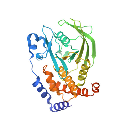
3QKP

Protein Tyrosine Phosphatase 1B - Apo W179F mutant with open WPD-loop


Experimental Data Snapshot

  • Method: X-RAY DIFFRACTION
  • Resolution: 2.05 Å
  • R-Value Free: 0.245 
  • R-Value Work: 0.204 
  • R-Value Observed: 0.206 

Starting Model: experimental
View more details

wwPDB Validation   3D Report Full Report


This is version 1.2 of the entry. See complete history


Literature

The molecular details of WPD-loop movement differ in the protein-tyrosine phosphatases YopH and PTP1B.

Brandao, T.A.Johnson, S.J.Hengge, A.C.

(2012) Arch Biochem Biophys 525: 53-59

  • DOI: https://doi.org/10.1016/j.abb.2012.06.002
  • Primary Citation of Related Structures:  
    3QKP, 3QKQ

  • PubMed Abstract: 

    The movement of a conserved protein loop (the WPD-loop) is important in catalysis by protein tyrosine phosphatases (PTPs). Using kinetics, isotope effects, and X-ray crystallography, the different effects arising from mutation of the conserved tryptophan in the WPD-loop were compared in two PTPs, the human PTP1B, and the bacterial YopH from Yersinia. Mutation of the conserved tryptophan in the WPD-loop to phenylalanine has a negligible effect on k(cat) in PTP1B and full loop movement is maintained. In contrast, the corresponding mutation in YopH reduces k(cat) by two orders of magnitude and the WPD loop locks in an intermediate position, disabling general acid catalysis. During loop movement the indole moiety of the WPD-loop tryptophan moves in opposite directions in the two enzymes. Comparisons of mammalian and bacterial PTPs reveal differences in the residues forming the hydrophobic pocket surrounding the conserved tryptophan. Thus, although WPD-loop movement is a conserved feature in PTPs, differences exist in the molecular details, and in the tolerance to mutation, in PTP1B compared to YopH. Despite high structural similarity of the active sites in both WPD-loop open and closed conformations, differences are identified in the molecular details associated with loop movement in PTPs from different organisms.


  • Organizational Affiliation

    Department of Chemistry and Biochemistry, Utah State University, Logan, UT 84322-0300, USA.


Macromolecules
Find similar proteins by:  (by identity cutoff)  |  3D Structure
Entity ID: 1
MoleculeChains Sequence LengthOrganismDetailsImage
Tyrosine-protein phosphatase non-receptor type 1321Homo sapiensMutation(s): 1 
Gene Names: ptp1bptpn1
EC: 3.1.3.48
UniProt & NIH Common Fund Data Resources
Find proteins for P18031 (Homo sapiens)
Explore P18031 
Go to UniProtKB:  P18031
PHAROS:  P18031
GTEx:  ENSG00000196396 
Entity Groups  
Sequence Clusters30% Identity50% Identity70% Identity90% Identity95% Identity100% Identity
UniProt GroupP18031
Sequence Annotations
Expand
  • Reference Sequence
Experimental Data & Validation

Experimental Data

  • Method: X-RAY DIFFRACTION
  • Resolution: 2.05 Å
  • R-Value Free: 0.245 
  • R-Value Work: 0.204 
  • R-Value Observed: 0.206 
  • Space Group: P 31 2 1
Unit Cell:
Length ( Å )Angle ( ˚ )
a = 88.282α = 90
b = 88.282β = 90
c = 104.535γ = 120
Software Package:
Software NamePurpose
DENZOdata reduction
SCALEPACKdata scaling
PHENIXrefinement
PDB_EXTRACTdata extraction
CrystalCleardata collection
HKL-2000data reduction
HKL-2000data scaling
PHASERphasing

Structure Validation

View Full Validation Report


Entry History 

Deposition Data

Revision History  (Full details and data files)

  • Version 1.0: 2012-08-08
    Type: Initial release
  • Version 1.1: 2017-11-08
    Changes: Refinement description
  • Version 1.2: 2023-09-13
    Changes: Data collection, Database references, Derived calculations, Refinement description