


Epidermin biosynthesis protein EpiD from Staphylococcus aureus.
Osipiuk, J., Makowska-Grzyska, M., Kwon, K., Anderson, W.F., Joachimiak, A.To be published.
Experimental Data Snapshot
Starting Model: experimental
View more details
Entity ID: 1 | |||||
|---|---|---|---|---|---|
| Molecule | Chains | Sequence Length | Organism | Details | Image |
| Epidermin biosynthesis protein EpiD | 175 | Staphylococcus aureus subsp. aureus COL | Mutation(s): 0 Gene Names: epiD, SACOL1875 | 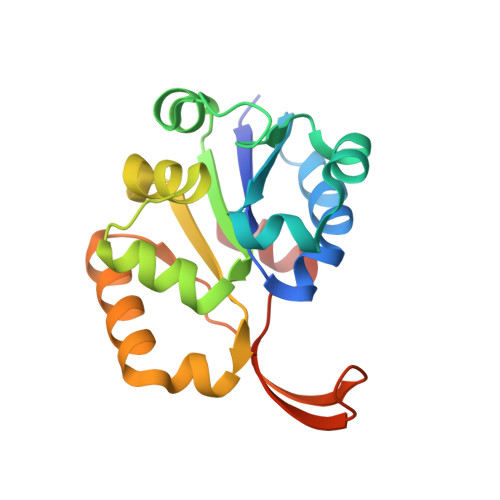 | |
UniProt | |||||
Find proteins for A0A0H2WWM9 (Staphylococcus aureus (strain COL)) Explore A0A0H2WWM9 Go to UniProtKB: A0A0H2WWM9 | |||||
Entity Groups | |||||
| Sequence Clusters | 30% Identity50% Identity70% Identity90% Identity95% Identity100% Identity | ||||
| UniProt Group | A0A0H2WWM9 | ||||
Sequence AnnotationsExpand | |||||
Reference Sequence | |||||
| Ligands 2 Unique | |||||
|---|---|---|---|---|---|
| ID | Chains | Name / Formula / InChI Key | 2D Diagram | 3D Interactions | |
| FMN Download:Ideal Coordinates CCD File | CA [auth K] DA [auth L] M [auth A] P [auth B] Q [auth C] | FLAVIN MONONUCLEOTIDE C17 H21 N4 O9 P FVTCRASFADXXNN-SCRDCRAPSA-N |  | ||
| CL Download:Ideal Coordinates CCD File | AA [auth J] BA [auth J] N [auth A] O [auth A] S [auth D] | CHLORIDE ION Cl VEXZGXHMUGYJMC-UHFFFAOYSA-M |  | ||
| Length ( Å ) | Angle ( ˚ ) |
|---|---|
| a = 160.991 | α = 90 |
| b = 111.256 | β = 97.25 |
| c = 154.569 | γ = 90 |
| Software Name | Purpose |
|---|---|
| DENZO | data reduction |
| SCALEPACK | data scaling |
| REFMAC | refinement |
| PDB_EXTRACT | data extraction |
| SBC-Collect | data collection |
| HKL-3000 | data reduction |
| HKL-3000 | data scaling |
| MOLREP | phasing |
| HKL-3000 | phasing |


