3QI5 | pdb_00003qi5

Crystal structure of human alkyladenine DNA glycosylase in complex with 3,N4-ethenocystosine containing duplex DNA


Experimental Data Snapshot

  • Method: X-RAY DIFFRACTION
  • Resolution: 2.20 Å
  • R-Value Free: 
    0.284 (Depositor), 0.307 (DCC) 
  • R-Value Work: 
    0.236 (Depositor) 
  • R-Value Observed: 
    0.239 (Depositor) 

Starting Model: experimental
View more details

wwPDB Validation 3D Report Full Report

Validation slider image for 3QI5

This is version 1.3 of the entry. See complete history

Literature

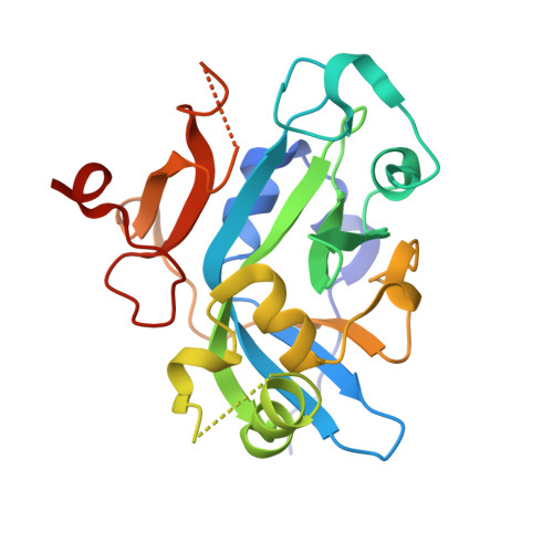
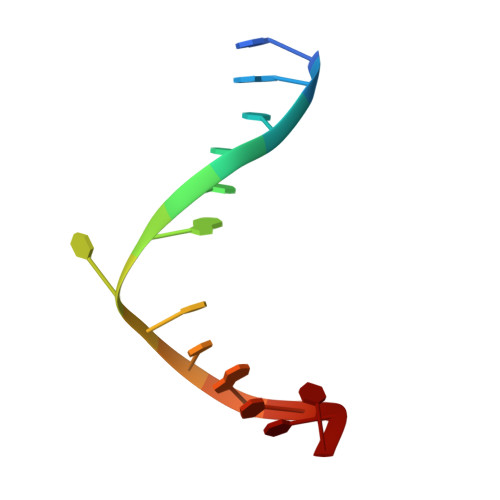
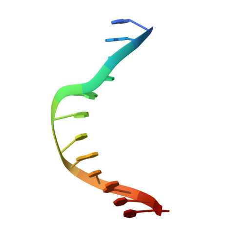
Structural Basis for the Inhibition of Human Alkyladenine DNA Glycosylase (AAG) by 3,N4-Ethenocytosine-containing DNA.

Lingaraju, G.M.Davis, C.A.Setser, J.W.Samson, L.D.Drennan, C.L.

(2011) J Biological Chem 286: 13205-13213

  • DOI: https://doi.org/10.1074/jbc.M110.192435
  • Primary Citation Related Structures: 
    3QI5

  • PubMed Abstract: 

    Reactive oxygen and nitrogen species, generated by neutrophils and macrophages in chronically inflamed tissues, readily damage DNA, producing a variety of potentially genotoxic etheno base lesions; such inflammation-related DNA damage is now known to contribute to carcinogenesis. Although the human alkyladenine DNA glycosylase (AAG) can specifically bind DNA containing either 1,N(6)-ethenoadenine (εA) lesions or 3,N(4)-ethenocytosine (εC) lesions, it can only excise εA lesions. AAG binds very tightly to DNA containing εC lesions, forming an abortive protein-DNA complex; such binding not only shields εC from repair by other enzymes but also inhibits AAG from acting on other DNA lesions. To understand the structural basis for inhibition, we have characterized the binding of AAG to DNA containing εC lesions and have solved a crystal structure of AAG bound to a DNA duplex containing the εC lesion. This study provides the first structure of a DNA glycosylase in complex with an inhibitory base lesion that is induced endogenously and that is also induced upon exposure to environmental agents such as vinyl chloride. We identify the primary cause of inhibition as a failure to activate the nucleotide base as an efficient leaving group and demonstrate that the higher binding affinity of AAG for εC versus εA is achieved through formation of an additional hydrogen bond between Asn-169 in the active site pocket and the O(2) of εC. This structure provides the basis for the design of AAG inhibitors currently being sought as an adjuvant for cancer chemotherapy.


  • Organizational Affiliation
    • Center for Environmental Health Sciences, Massachusetts Institute of Technology, Cambridge, Massachusetts 02139, USA.

Macromolecule Content 

  • Total Structure Weight: 64.67 kDa 
  • Atom Count: 4,516 
  • Modeled Residue Count: 468 
  • Deposited Residue Count: 490 
  • Unique protein chains: 1
  • Unique nucleic acid chains: 2

Macromolecules


Find similar proteins by:|  3D Structure
Entity ID: 1
MoleculeChains  Sequence LengthOrganismDetailsImage
DNA-3-methyladenine glycosylase
A, B
219Homo sapiensMutation(s): 0 
Gene Names: MPGAAGANPGMID1
EC: 3.2.2.21
UniProt & NIH Common Fund Data Resources
Find proteins for P29372 (Homo sapiens)
Explore P29372 
Go to UniProtKB:  P29372
PHAROS:  P29372
GTEx:  ENSG00000103152 
Entity Groups
Sequence Clusters30% Identity50% Identity70% Identity90% Identity95% Identity100% Identity
UniProt GroupP29372
Sequence Annotations
Expand
Reference Sequence
Find similar nucleic acids by:  Sequence
Entity ID: 2
MoleculeChains LengthOrganismImage
DNA (5'-D(*GP*AP*CP*AP*TP*GP*(EDC)P*TP*TP*GP*CP*CP*T)-3')
C, E
13N/A
Sequence Annotations
Expand
Reference Sequence
Find similar nucleic acids by:  Sequence
Entity ID: 3
MoleculeChains LengthOrganismImage
DNA (5'-D(*GP*GP*CP*AP*AP*GP*CP*AP*TP*GP*TP*CP*A)-3')
D, F
13N/A
Sequence Annotations
Expand
Reference Sequence

Experimental Data & Validation

Experimental Data

  • Method: X-RAY DIFFRACTION
  • Resolution: 2.20 Å
  • R-Value Free:  0.284 (Depositor), 0.307 (DCC) 
  • R-Value Work:  0.236 (Depositor) 
  • R-Value Observed: 0.239 (Depositor) 
Space Group: P 1
Unit Cell:
Length ( Å )Angle ( ˚ )
a = 41.226α = 81.23
b = 41.22β = 88.4
c = 82.144γ = 89.15
Software Package:
Software NamePurpose
ADSCdata collection
PHASERphasing
REFMACrefinement
DENZOdata reduction
SCALEPACKdata scaling

Structure Validation

View Full Validation Report


Entry History 

Deposition Data

Revision History  (Full details and data files)

  • Version 1.0: 2011-03-09
    Type: Initial release
  • Version 1.1: 2011-07-13
    Changes: Version format compliance
  • Version 1.2: 2012-01-18
    Changes: Atomic model
  • Version 1.3: 2023-09-13
    Changes: Data collection, Database references, Derived calculations, Refinement description