3QE7 | pdb_00003qe7

Crystal Structure of Uracil Transporter--UraA


Experimental Data Snapshot

  • Method: X-RAY DIFFRACTION
  • Resolution: 2.78 Å
  • R-Value Free: 
    0.303 (Depositor), 0.312 (DCC) 
  • R-Value Work: 
    0.248 (Depositor), 0.257 (DCC) 
  • R-Value Observed: 
    0.251 (Depositor) 

wwPDB Validation 3D Report Full Report

Validation slider image for 3QE7

Ligand Structure Quality Assessment 


This is version 1.3 of the entry. See complete history

Literature

Structure and mechanism of the uracil transporter UraA

Lu, F.Li, S.Jiang, Y.Jiang, J.Fan, H.Lu, G.Deng, D.Dang, S.Zhang, X.Wang, J.Yan, N.

(2011) Nature 472: 243-246

  • DOI: https://doi.org/10.1038/nature09885
  • Primary Citation Related Structures: 
    3QE7

  • PubMed Abstract: 

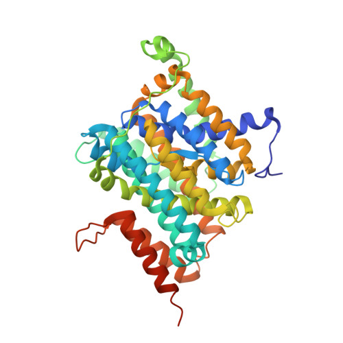
    The nucleobase/ascorbate transporter (NAT) proteins, also known as nucleobase/cation symporter 2 (NCS2) proteins, are responsible for the uptake of nucleobases in all kingdoms of life and for the transport of vitamin C in mammals. Despite functional characterization of the NAT family members in bacteria, fungi and mammals, detailed structural information remains unavailable. Here we report the crystal structure of a representative NAT protein, the Escherichia coli uracil/H(+) symporter UraA, in complex with uracil at a resolution of 2.8 Å. UraA has a novel structural fold, with 14 transmembrane segments (TMs) divided into two inverted repeats. A pair of antiparallel β-strands is located between TM3 and TM10 and has an important role in structural organization and substrate recognition. The structure is spatially arranged into a core domain and a gate domain. Uracil, located at the interface between the two domains, is coordinated mainly by residues from the core domain. Structural analysis suggests that alternating access of the substrate may be achieved through conformational changes of the gate domain.


  • Organizational Affiliation
    • State Key Laboratory of Bio-membrane and Membrane Biotechnology, Center for Structural Biology, School of Life Sciences, Tsinghua University, Beijing 100084, China.

Macromolecule Content 

  • Total Structure Weight: 45.51 kDa 
  • Atom Count: 3,030 
  • Modeled Residue Count: 407 
  • Deposited Residue Count: 429 
  • Unique protein chains: 1

Macromolecules

Find similar proteins by:|  3D Structure
Entity ID: 1
MoleculeChains  Sequence LengthOrganismDetailsImage
Uracil permease429Escherichia coli K-12Mutation(s): 0 
Gene Names: uraAb2497JW2482
Membrane Entity: Yes 
UniProt
Find proteins for P0AGM7 (Escherichia coli (strain K12))
Explore P0AGM7 
Go to UniProtKB:  P0AGM7
Entity Groups
Sequence Clusters30% Identity50% Identity70% Identity90% Identity95% Identity100% Identity
UniProt GroupP0AGM7
Sequence Annotations
Expand
Reference Sequence

Small Molecules

Experimental Data & Validation

Experimental Data

  • Method: X-RAY DIFFRACTION
  • Resolution: 2.78 Å
  • R-Value Free:  0.303 (Depositor), 0.312 (DCC) 
  • R-Value Work:  0.248 (Depositor), 0.257 (DCC) 
  • R-Value Observed: 0.251 (Depositor) 
Space Group: P 64 2 2
Unit Cell:
Length ( Å )Angle ( ˚ )
a = 96.76α = 90
b = 96.76β = 90
c = 251.95γ = 120
Software Package:
Software NamePurpose
BSSdata collection
PHASERphasing
PHENIXrefinement
DENZOdata reduction
SCALEPACKdata scaling

Structure Validation

View Full Validation Report


Ligand Structure Quality Assessment 


Entry History 

Deposition Data

Revision History  (Full details and data files)

  • Version 1.0: 2011-03-23
    Type: Initial release
  • Version 1.1: 2011-07-13
    Changes: Version format compliance
  • Version 1.2: 2020-07-29
    Type: Remediation
    Reason: Carbohydrate remediation
    Changes: Data collection, Derived calculations, Structure summary
  • Version 1.3: 2024-03-20
    Changes: Data collection, Database references, Structure summary