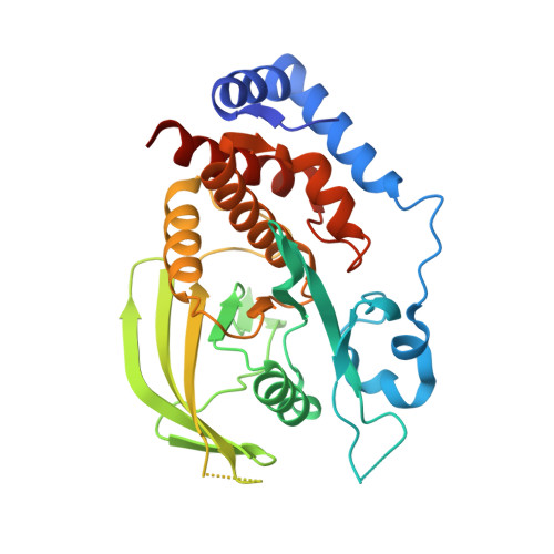
Small molecule receptor protein tyrosine phosphatase [gamma](RPTP[gamma]) ligands that inhibit phosphatase activity via perturbation of the tryptophan-proline-aspartate (WPD) loop
Sheriff, S., Beno, B.R., Zhai, W., Kostich, W.A., McDonnell, P.A., Kish, K., Goldfarb, V., Gao, M., Kiefer, S.E., Yanchunas, J., Huang, Y., Shi, S., Zhu, S., Dzierba, C., Bronson, J., Macor, J.E., Appiah, K.K., Westphal, R.S., O'Connell, J., Gerritz, S.W.(2011) J Med Chem 54: 6548-6562
- PubMed: 21882820
- DOI: https://doi.org/10.1021/jm2003766
- Primary Citation of Related Structures:
3QCB, 3QCC, 3QCD, 3QCE, 3QCF, 3QCG, 3QCH, 3QCI, 3QCJ, 3QCK, 3QCL, 3QCM, 3QCN - PubMed Abstract:
Protein tyrosine phosphatases (PTPs) catalyze the dephosphorylation of tyrosine residues, a process that involves a conserved tryptophan-proline-aspartate (WPD) loop in catalysis. In previously determined structures of PTPs, the WPD-loop has been observed in either an "open" conformation or a "closed" conformation. In the current work, X-ray structures of the catalytic domain of receptor-like protein tyrosine phosphatase γ (RPTPγ) revealed a ligand-induced "superopen" conformation not previously reported for PTPs. In the superopen conformation, the ligand acts as an apparent competitive inhibitor and binds in a small hydrophobic pocket adjacent to, but distinct from, the active site. In the open and closed WPD-loop conformations of RPTPγ, the side chain of Trp1026 partially occupies this pocket. In the superopen conformation, Trp1026 is displaced allowing a 3,4-dichlorobenzyl substituent to occupy this site. The bound ligand prevents closure of the WPD-loop over the active site and disrupts the catalytic cycle of the enzyme.
- Bristol-Myers Squibb Research and Development, P.O. Box 4000, Princeton, New Jersey 08543-4000, United States. steven.sheriff@bms.com
Organizational Affiliation:


