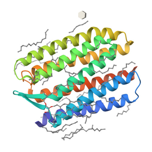
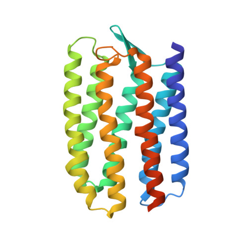
3QAP

Crystal structure of Natronomonas pharaonis sensory rhodopsin II in the ground state


Experimental Data Snapshot

  • Method: X-RAY DIFFRACTION
  • Resolution: 1.90 Å
  • R-Value Free: 0.173 
  • R-Value Work: 0.152 
  • R-Value Observed: 0.153 

wwPDB Validation   3D Report Full Report


Ligand Structure Quality Assessment 

Created with Raphaël 2.3.0Worse 01 BetterLigand structure goodness of fit to experimental dataBest fitted L2PClick on this verticalbar to view detailsBest fitted BOGClick on this verticalbar to view detailsBest fitted RETClick on this verticalbar to view detailsBest fitted LFAClick on this verticalbar to view details

This is version 1.3 of the entry. See complete history


Literature

Active State of Sensory Rhodopsin II: Structural Determinants for Signal Transfer and Proton Pumping.

Gushchin, I.Reshetnyak, A.Borshchevskiy, V.Ishchenko, A.Round, E.Grudinin, S.Engelhard, M.Buldt, G.Gordeliy, V.

(2011) J Mol Biol 412: 591-600

  • DOI: https://doi.org/10.1016/j.jmb.2011.07.022
  • Primary Citation of Related Structures:  
    3QAP, 3QDC

  • PubMed Abstract: 

    The molecular mechanism of transmembrane signal transduction is still a pertinent question in cellular biology. Generally, a receptor can transfer an external signal via its cytoplasmic surface, as found for G-protein-coupled receptors such as rhodopsin, or via the membrane domain, such as that in sensory rhodopsin II (SRII) in complex with its transducer, HtrII. In the absence of HtrII, SRII functions as a proton pump. Here, we report on the crystal structure of the active state of uncomplexed SRII from Natronomonas pharaonis, NpSRII. The problem with a dramatic loss of diffraction quality upon loading of the active state was overcome by growing better crystals and by reducing the occupancy of the state. The conformational changes in the region comprising helices F and G are similar to those observed for the NpSRII-transducer complex but are much more pronounced. The meaning of these differences for the understanding of proton pumping and signal transduction by NpSRII is discussed.


  • Organizational Affiliation

    Laboratoire des Protéines Membranaires, Institut de Biologie Structurale J.-P. Ebel, UMR5075 CEA-CNRS-UJF, 38027 Grenoble, France.


Macromolecules
Find similar proteins by:  (by identity cutoff)  |  3D Structure
Entity ID: 1
MoleculeChains Sequence LengthOrganismDetailsImage
Sensory rhodopsin-2239Natronomonas pharaonisMutation(s): 0 
Gene Names: sop2sopII
Membrane Entity: Yes 
UniProt
Find proteins for P42196 (Natronomonas pharaonis)
Explore P42196 
Go to UniProtKB:  P42196
Entity Groups  
Sequence Clusters30% Identity50% Identity70% Identity90% Identity95% Identity100% Identity
UniProt GroupP42196
Sequence Annotations
Expand
  • Reference Sequence
Small Molecules
Ligands 4 Unique
IDChains Name / Formula / InChI Key2D Diagram3D Interactions
L2P
Query on L2P

Download Ideal Coordinates CCD File 
S [auth A],
V [auth A]
2,3-DI-PHYTANYL-GLYCEROL
C43 H88 O3
ISDBCJSGCHUHFI-UMZPFTBHSA-N
BOG
Query on BOG

Download Ideal Coordinates CCD File 
C [auth A]octyl beta-D-glucopyranoside
C14 H28 O6
HEGSGKPQLMEBJL-RKQHYHRCSA-N
RET
Query on RET

Download Ideal Coordinates CCD File 
B [auth A]RETINAL
C20 H28 O
NCYCYZXNIZJOKI-OVSJKPMPSA-N
LFA
Query on LFA

Download Ideal Coordinates CCD File 
AA [auth A]
BA [auth A]
CA [auth A]
D [auth A]
DA [auth A]
EICOSANE
C20 H42
CBFCDTFDPHXCNY-UHFFFAOYSA-N
Experimental Data & Validation

Experimental Data

  • Method: X-RAY DIFFRACTION
  • Resolution: 1.90 Å
  • R-Value Free: 0.173 
  • R-Value Work: 0.152 
  • R-Value Observed: 0.153 
  • Space Group: C 2 2 21
Unit Cell:
Length ( Å )Angle ( ˚ )
a = 86.99α = 90
b = 128.09β = 90
c = 50.63γ = 90
Software Package:
Software NamePurpose
ADSCdata collection
MOLREPphasing
REFMACrefinement
MOSFLMdata reduction
SCALAdata scaling

Structure Validation

View Full Validation Report


Ligand Structure Quality Assessment 

Created with Raphaël 2.3.0Worse 01 BetterLigand structure goodness of fit to experimental dataBest fitted L2PClick on this verticalbar to view detailsBest fitted BOGClick on this verticalbar to view detailsBest fitted RETClick on this verticalbar to view detailsBest fitted LFAClick on this verticalbar to view details

Entry History 

Deposition Data

Revision History  (Full details and data files)

  • Version 1.0: 2011-09-14
    Type: Initial release
  • Version 1.1: 2011-09-28
    Changes: Database references
  • Version 1.2: 2020-07-29
    Type: Remediation
    Reason: Carbohydrate remediation
    Changes: Advisory, Data collection, Derived calculations, Structure summary
  • Version 1.3: 2024-02-21
    Changes: Advisory, Data collection, Database references, Structure summary