3QA3 | pdb_00003qa3

Crystal Structure of A-domain in complex with antibody


Experimental Data Snapshot

  • Method: X-RAY DIFFRACTION
  • Resolution: 3.00 Å
  • R-Value Free: 
    0.224 (Depositor), 0.229 (DCC) 
  • R-Value Work: 
    0.194 (Depositor), 0.201 (DCC) 
  • R-Value Observed: 
    0.196 (Depositor) 

Starting Model: experimental
View more details

wwPDB Validation 3D Report Full Report

Validation slider image for 3QA3

This is version 1.3 of the entry. See complete history

Literature

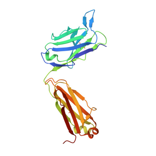
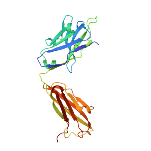
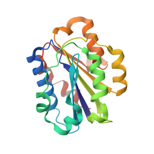
Stable Coordination of the Inhibitory Ca2+ Ion at the Metal Ion-Dependent Adhesion Site in Integrin CD11b/CD18 by an Antibody-Derived Ligand Aspartate: Implications for Integrin Regulation and Structure-Based Drug Design.

Mahalingam, B.Ajroud, K.Alonso, J.L.Anand, S.Adair, B.D.Horenstein, A.L.Malavasi, F.Xiong, J.P.Arnaout, M.A.

(2011) J Immunol 187: 6393-6401

  • DOI: https://doi.org/10.4049/jimmunol.1102394
  • Primary Citation Related Structures: 
    3Q3G, 3QA3

  • PubMed Abstract: 

    A central feature of integrin interaction with physiologic ligands is the monodentate binding of a ligand carboxylate to a Mg(2+) ion hexacoordinated at the metal ion-dependent adhesion site (MIDAS) in the integrin A domain. This interaction stabilizes the A domain in the high-affinity state, which is distinguished from the default low-affinity state by tertiary changes in the domain that culminate in cell adhesion. Small molecule ligand-mimetic integrin antagonists act as partial agonists, eliciting similar activating conformational changes in the A domain, which has contributed to paradoxical adhesion and increased patient mortality in large clinical trials. As with other ligand-mimetic integrin antagonists, the function-blocking mAb 107 binds MIDAS of integrin CD11b/CD18 A domain (CD11bA), but in contrast, it favors the inhibitory Ca(2+) ion over the Mg(2+) ion at MIDAS. We determined the crystal structures of the Fab fragment of mAb 107 complexed to the low- and high-affinity states of CD11bA. Favored binding of the Ca(2+) ion at MIDAS is caused by the unusual symmetric bidentate ligation of a Fab-derived ligand Asp to a heptacoordinated MIDAS Ca(2+) ion. Binding of the Fab fragment of mAb 107 to CD11bA did not trigger the activating tertiary changes in the domain or in the full-length integrin. These data show that the denticity of the ligand Asp/Glu can modify the divalent cation selectivity at MIDAS and hence integrin function. Stabilizing the Ca(2+) ion at MIDAS by bidentate ligation to a ligand Asp/Glu may provide one approach for designing pure integrin antagonists.


  • Organizational Affiliation
    • Structural Biology Program, Division of Nephrology, Massachusetts General Hospital, Harvard Medical School, Charlestown, MA 02129, USA.

Macromolecule Content 

  • Total Structure Weight: 282.01 kDa 
  • Atom Count: 19,772 
  • Modeled Residue Count: 2,519 
  • Deposited Residue Count: 2,536 
  • Unique protein chains: 3

Macromolecules

Find similar proteins by:|  3D Structure
Entity ID: 1
MoleculeChains  Sequence LengthOrganismDetailsImage
Antibody Light chainA [auth C],
D [auth A],
G [auth F],
J
220Mus musculusMutation(s): 0 
Find similar proteins by:|  3D Structure
Entity ID: 2
MoleculeChains  Sequence LengthOrganismDetailsImage
Antibody Heavy chainB [auth D],
E [auth B],
H,
K
224Mus musculusMutation(s): 0 
Find similar proteins by:|  3D Structure
Entity ID: 3
MoleculeChains  Sequence LengthOrganismDetailsImage
Integrin alpha-MC [auth G],
F [auth E],
I,
L
190Homo sapiensMutation(s): 1 
Gene Names: ITGAMCD11BCR3A
UniProt & NIH Common Fund Data Resources
Find proteins for P11215 (Homo sapiens)
Explore P11215 
Go to UniProtKB:  P11215
PHAROS:  P11215
GTEx:  ENSG00000169896 
Entity Groups
Sequence Clusters30% Identity50% Identity70% Identity90% Identity95% Identity100% Identity
UniProt GroupP11215
Sequence Annotations
Expand
Reference Sequence

Small Molecules

Ligands 3 Unique
IDChains Name / Formula / InChI Key2D Diagram3D Interactions
GOL

Query on GOL


Download:Ideal Coordinates CCD File
O [auth D],
S [auth G],
T [auth G],
U [auth A],
V [auth B]
GLYCEROL
C3 H8 O3
PEDCQBHIVMGVHV-UHFFFAOYSA-N
EDO

Query on EDO


Download:Ideal Coordinates CCD File
AA [auth J]
BA [auth K]
M [auth C]
N [auth C]
P [auth D]
AA [auth J],
BA [auth K],
M [auth C],
N [auth C],
P [auth D],
Q [auth D],
X [auth H],
Z [auth J]
1,2-ETHANEDIOL
C2 H6 O2
LYCAIKOWRPUZTN-UHFFFAOYSA-N
CA

Query on CA


Download:Ideal Coordinates CCD File
CA [auth L],
R [auth G],
W [auth E],
Y [auth I]
CALCIUM ION
Ca
BHPQYMZQTOCNFJ-UHFFFAOYSA-N

Experimental Data & Validation

Experimental Data

  • Method: X-RAY DIFFRACTION
  • Resolution: 3.00 Å
  • R-Value Free:  0.224 (Depositor), 0.229 (DCC) 
  • R-Value Work:  0.194 (Depositor), 0.201 (DCC) 
  • R-Value Observed: 0.196 (Depositor) 
Space Group: P 21 21 21
Unit Cell:
Length ( Å )Angle ( ˚ )
a = 82.604α = 90
b = 158.346β = 90
c = 233.319γ = 90
Software Package:
Software NamePurpose
BUSTERrefinement
HKL-2000data reduction
HKL-2000data scaling
PHASERphasing

Structure Validation

View Full Validation Report


Entry History 

Deposition Data

Revision History  (Full details and data files)

  • Version 1.0: 2011-11-30
    Type: Initial release
  • Version 1.1: 2011-12-28
    Changes: Database references
  • Version 1.2: 2023-09-13
    Changes: Data collection, Database references, Derived calculations, Refinement description
  • Version 1.3: 2024-10-09
    Changes: Structure summary