3Q8T | pdb_00003q8t

Crystal structure of the coiled coil domain of Beclin 1, an essential autophagy protein


Experimental Data Snapshot

  • Method: X-RAY DIFFRACTION
  • Resolution: 1.90 Å
  • R-Value Free: 
    0.236 (Depositor), 0.235 (DCC) 
  • R-Value Work: 
    0.209 (Depositor), 0.208 (DCC) 
  • R-Value Observed: 
    0.210 (Depositor) 

wwPDB Validation 3D Report Full Report

Validation slider image for 3Q8T

This is version 1.2 of the entry. See complete history

Literature

Imperfect interface of Beclin1 coiled-coil domain regulates homodimer and heterodimer formation with Atg14L and UVRAG

Li, X.He, L.Che, K.H.Funderburk, S.F.Pan, L.Pan, N.Zhang, M.Yue, Z.Zhao, Y.

(2012) Nat Commun 3: 662-662

  • DOI: https://doi.org/10.1038/ncomms1648
  • Primary Citation Related Structures: 
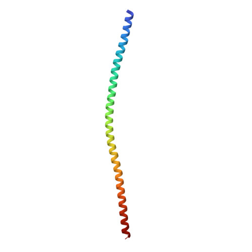
    3Q8T

  • PubMed Abstract: 

    Beclin 1 is a core component of the Class III Phosphatidylinositol 3-Kinase VPS34 complex. The coiled coil domain of Beclin 1 serves as an interaction platform for assembly of distinct Atg14L- and UVRAG-containing complexes to modulate VPS34 activity. Here we report the crystal structure of the coiled coil domain that forms an antiparallel dimer and is rendered metastable by a series of 'imperfect' a-d' pairings at its coiled coil interface. Atg14L and UVRAG promote the transition of metastable homodimeric Beclin 1 to heterodimeric Beclin1-Atg14L/UVRAG assembly. Beclin 1 mutants with their 'imperfect' a-d' pairings modified to enhance self-interaction, show distinctively altered interactions with Atg14L or UVRAG. These results suggest that specific utilization of the dimer interface and modulation of the homodimer-heterodimer transition by Beclin 1-interacting partners may underlie the molecular mechanism that controls the formation of various Beclin1-VPS34 subcomplexes to exert their effect on an array of VPS34-related activities, including autophagy.


  • Organizational Affiliation
    • Department of Applied Biology and Chemical Technology, State Key Laboratory of Chirosciences, The Hong Kong Polytechnic University, Hung Hom, Kowloon, Hong Kong, PR China.

Macromolecule Content 

  • Total Structure Weight: 23.06 kDa 
  • Atom Count: 1,871 
  • Modeled Residue Count: 189 
  • Deposited Residue Count: 192 
  • Unique protein chains: 1

Macromolecules

Find similar proteins by:|  3D Structure
Entity ID: 1
MoleculeChains  Sequence LengthOrganismDetailsImage
Beclin-1
A, B
96Rattus norvegicusMutation(s): 0 
Gene Names: Becn1
UniProt
Find proteins for Q91XJ1 (Rattus norvegicus)
Explore Q91XJ1 
Go to UniProtKB:  Q91XJ1
Entity Groups
Sequence Clusters30% Identity50% Identity70% Identity90% Identity95% Identity100% Identity
UniProt GroupQ91XJ1
Sequence Annotations
Expand
Reference Sequence

Experimental Data & Validation

Experimental Data

  • Method: X-RAY DIFFRACTION
  • Resolution: 1.90 Å
  • R-Value Free:  0.236 (Depositor), 0.235 (DCC) 
  • R-Value Work:  0.209 (Depositor), 0.208 (DCC) 
  • R-Value Observed: 0.210 (Depositor) 
Space Group: C 1 2 1
Unit Cell:
Length ( Å )Angle ( ˚ )
a = 103.78α = 90
b = 38.35β = 97.92
c = 65.75γ = 90
Software Package:
Software NamePurpose
CrystalCleardata collection
MLPHAREphasing
REFMACrefinement
d*TREKdata reduction
d*TREKdata scaling

Structure Validation

View Full Validation Report


Entry History 

Deposition Data

Revision History  (Full details and data files)

  • Version 1.0: 2012-01-18
    Type: Initial release
  • Version 1.1: 2012-02-22
    Changes: Database references
  • Version 1.2: 2024-03-20
    Changes: Data collection, Database references