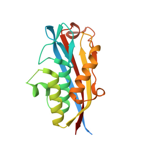
3Q8H

Crystal structure of 2c-methyl-d-erythritol 2,4-cyclodiphosphate synthase from burkholderia pseudomallei in complex with cytidine derivative EBSI01028


Experimental Data Snapshot

  • Method: X-RAY DIFFRACTION
  • Resolution: 1.75 Å
  • R-Value Free: 0.184 
  • R-Value Work: 0.155 
  • R-Value Observed: 0.156 

Starting Model: experimental
View more details

wwPDB Validation   3D Report Full Report


Ligand Structure Quality Assessment 


This is version 1.4 of the entry. See complete history


Literature

Cytidine derivatives as IspF inhibitors of Burkolderia pseudomallei.

Zhang, Z.Jakkaraju, S.Blain, J.Gogol, K.Zhao, L.Hartley, R.C.Karlsson, C.A.Staker, B.L.Edwards, T.E.Stewart, L.J.Myler, P.J.Clare, M.Begley, D.W.Horn, J.R.Hagen, T.J.

(2013) Bioorg Med Chem Lett 23: 6860-6863

  • DOI: https://doi.org/10.1016/j.bmcl.2013.09.101
  • Primary Citation of Related Structures:  
    3KE1, 3Q8H

  • PubMed Abstract: 

    Published biological data suggest that the methyl erythritol phosphate (MEP) pathway, a non-mevalonate isoprenoid biosynthetic pathway, is essential for certain bacteria and other infectious disease organisms. One highly conserved enzyme in the MEP pathway is 2C-methyl-d-erythritol 2,4-cyclodiphosphate synthase (IspF). Fragment-bound complexes of IspF from Burkholderia pseudomallei were used to design and synthesize a series of molecules linking the cytidine moiety to different zinc pocket fragment binders. Testing by surface plasmon resonance (SPR) found one molecule in the series to possess binding affinity equal to that of cytidine diphosphate, despite lacking any metal-coordinating phosphate groups. Close inspection of the SPR data suggest different binding stoichiometries between IspF and test compounds. Crystallographic analysis shows important variations between the binding mode of one synthesized compound and the pose of the bound fragment from which it was designed. The binding modes of these molecules add to our structural knowledge base for IspF and suggest future refinements in this compound series.


  • Organizational Affiliation

    Department of Chemistry and Biochemistry, Northern Illinois University, DeKalb, IL, USA.


Macromolecules
Find similar proteins by:  (by identity cutoff)  |  3D Structure
Entity ID: 1
MoleculeChains Sequence LengthOrganismDetailsImage
2-C-methyl-D-erythritol 2,4-cyclodiphosphate synthase
A, B, C
183Burkholderia pseudomalleiMutation(s): 0 
Gene Names: ispFmecSBPSL2098
EC: 4.6.1.12
UniProt
Find proteins for Q63T71 (Burkholderia pseudomallei (strain K96243))
Explore Q63T71 
Go to UniProtKB:  Q63T71
Entity Groups  
Sequence Clusters30% Identity50% Identity70% Identity90% Identity95% Identity100% Identity
UniProt GroupQ63T71
Sequence Annotations
Expand
  • Reference Sequence
Small Molecules
Ligands 4 Unique
IDChains Name / Formula / InChI Key2D Diagram3D Interactions
AO9
Query on AO9

Download Ideal Coordinates CCD File 
E [auth A],
I [auth A],
K [auth B]
5'-deoxy-5'-[(imidazo[2,1-b][1,3]thiazol-5-ylcarbonyl)amino]cytidine
C15 H16 N6 O5 S
NPCKGXDYODQBDY-UORFTKCHSA-N
ZN
Query on ZN

Download Ideal Coordinates CCD File 
D [auth A],
J [auth B],
L [auth C]
ZINC ION
Zn
PTFCDOFLOPIGGS-UHFFFAOYSA-N
EDO
Query on EDO

Download Ideal Coordinates CCD File 
G [auth A],
H [auth A]
1,2-ETHANEDIOL
C2 H6 O2
LYCAIKOWRPUZTN-UHFFFAOYSA-N
K
Query on K

Download Ideal Coordinates CCD File 
F [auth A]POTASSIUM ION
K
NPYPAHLBTDXSSS-UHFFFAOYSA-N
Binding Affinity Annotations 
IDSourceBinding Affinity
AO9 PDBBind:  3Q8H Kd: 7.00e+4 (nM) from 1 assay(s)
Experimental Data & Validation

Experimental Data

  • Method: X-RAY DIFFRACTION
  • Resolution: 1.75 Å
  • R-Value Free: 0.184 
  • R-Value Work: 0.155 
  • R-Value Observed: 0.156 
  • Space Group: C 1 2 1
Unit Cell:
Length ( Å )Angle ( ˚ )
a = 117.69α = 90
b = 67.76β = 96.02
c = 60.02γ = 90
Software Package:
Software NamePurpose
XSCALEdata scaling
PHASERphasing
REFMACrefinement
PDB_EXTRACTdata extraction

Structure Validation

View Full Validation Report


Ligand Structure Quality Assessment 


Entry History 

Deposition Data

Revision History  (Full details and data files)

  • Version 1.0: 2011-01-19
    Type: Initial release
  • Version 1.1: 2011-07-13
    Changes: Version format compliance
  • Version 1.2: 2013-10-30
    Changes: Database references
  • Version 1.3: 2013-12-11
    Changes: Database references
  • Version 1.4: 2023-09-13
    Changes: Data collection, Database references, Derived calculations, Refinement description