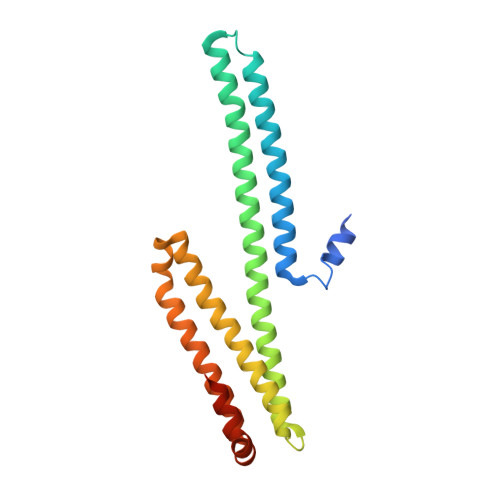
Crystal Structure of the E2 Domain of Amyloid Precursor Protein-like Protein 1 in Complex with Sucrose Octasulfate.
Xue, Y., Lee, S., Wang, Y., Ha, Y.(2011) J Biological Chem 286: 29748-29757
- PubMed: 21715329
- DOI: https://doi.org/10.1074/jbc.M111.219659
- Primary Citation of Related Structures:
3Q7G, 3Q7L - PubMed Abstract:
Missense mutations in the amyloid precursor protein (APP) gene can cause familial Alzheimer disease. It is thought that APP and APP-like proteins (APLPs) may play a role in adhesion and signal transduction because their ectodomains interact with components of the extracellular matrix. Heparin binding induces dimerization of APP and APLPs. To help explain how these proteins interact with heparin, we have determined the crystal structure of the E2 domain of APLP1 in complex with sucrose octasulfate (SOS). A total of three SOS molecules are bound to the E2 dimer. Two SOSs are bound inside a narrow intersubdomain groove, and the third SOS is bound near the two-fold axis of the protein. Mutational analyses show that most residues interacting with SOS also contribute to heparin binding, although in varying degrees; a deep pocket, defined by His-376, Lys-422, and Arg-429, and an interfacial site between Lys-314 and its symmetry mate are most important in the binding of the negatively charged polysaccharide. Comparison with a lower resolution APP structure shows that all key heparin binding residues are conserved and identically positioned, suggesting that APLP1 and APP may bind heparin similarly. In transfected HEK-293 cells, mutating residues responsible for heparin binding causes little change in the proteolysis of APP by the secretases. However, mutating a pair of conserved basic residues (equivalent to Arg-414 and Arg-415 of APLP1) immediately adjacent to the heparin binding site affects both the maturation and the processing of APP.
- Department of Pharmacology, Yale School of Medicine, New Haven, Connecticut 06520, USA.
Organizational Affiliation:


